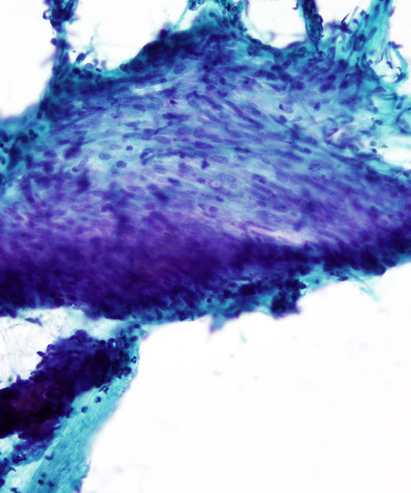
Medium power: Pap stain
Small fragment of bland spindle cells
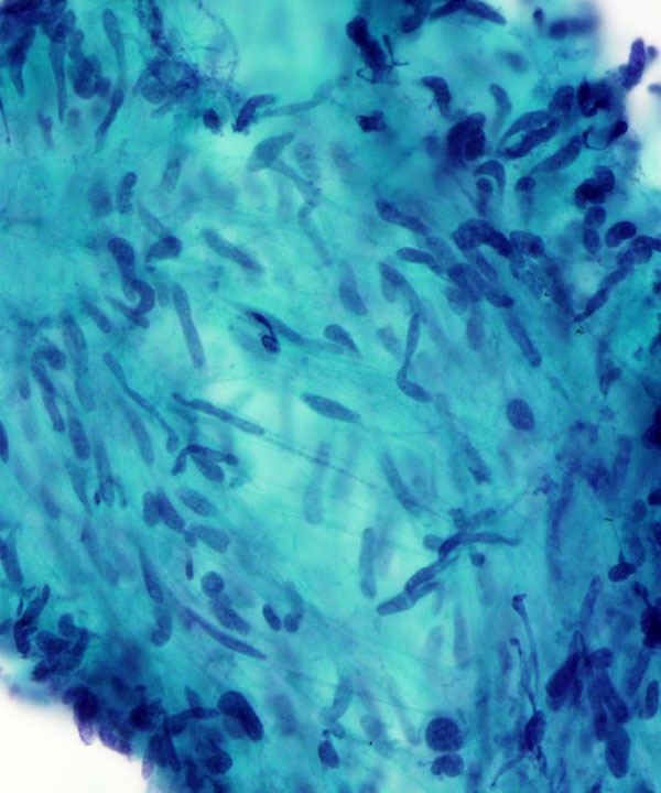
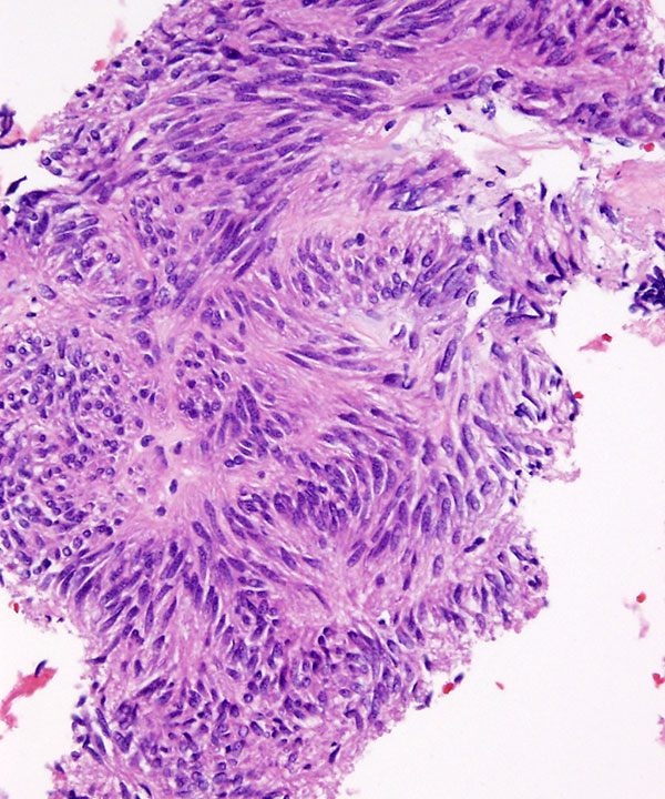
Medium power: Pap stain
Fragment of bland spindle cells with uniform elongated nuclei
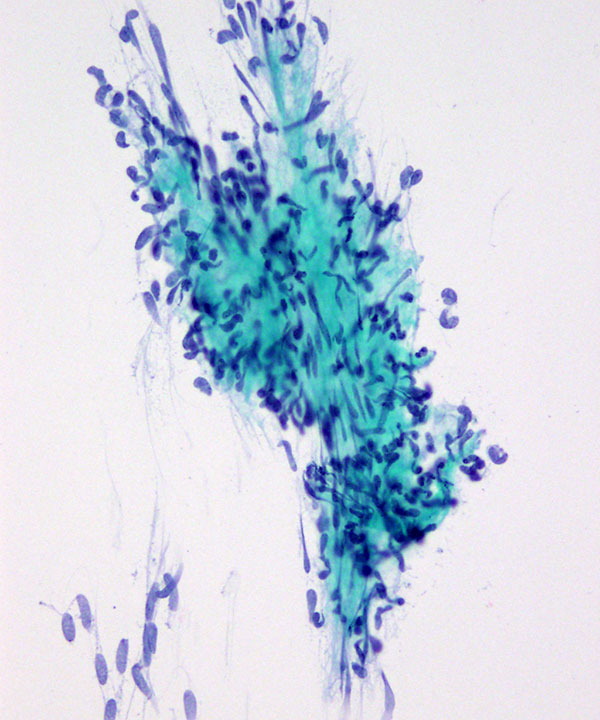
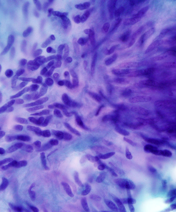
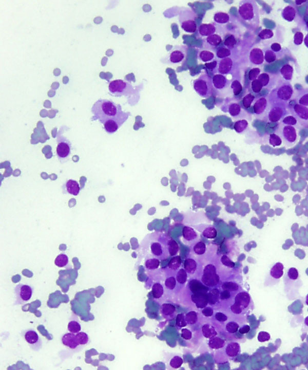
Medium power: Pap stain
Crowded aggregate of bland spindle cells with uniform elongated nuclei
High power: Pap stain
Bland spindle cells with uniform elongated nuclei, fine chromatin and delicate cytoplasm
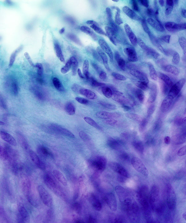
High power: Pap stain
Spindle cells with uniform elongated nuclei, fine chromatin and delicate cytoplasm
Cell block: H&E stain
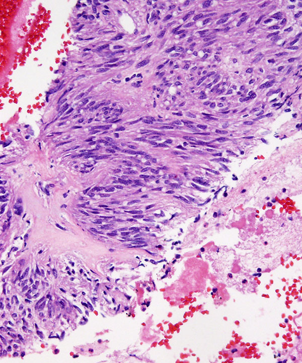
Cell block: H&E stain
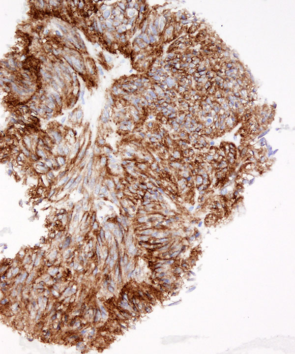
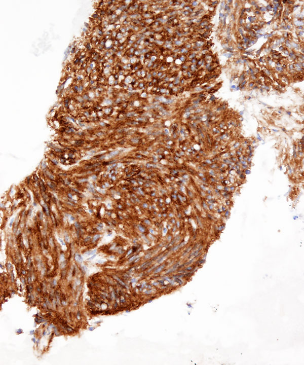
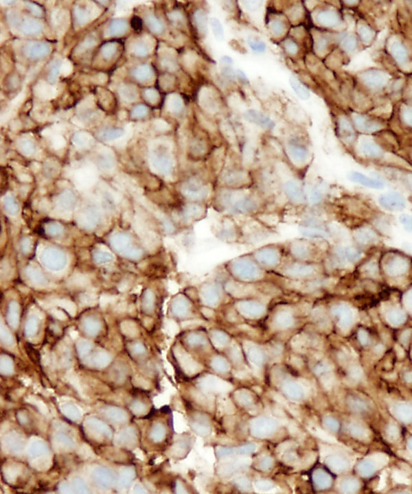
Cell block: IHC for CD117
Cell block: IHC for DOG-1
Medium power: DQ stain
Epithelioid variant GIST
Polygonal cells with round to oval nuclei, and moderate to abundant cytoplasm
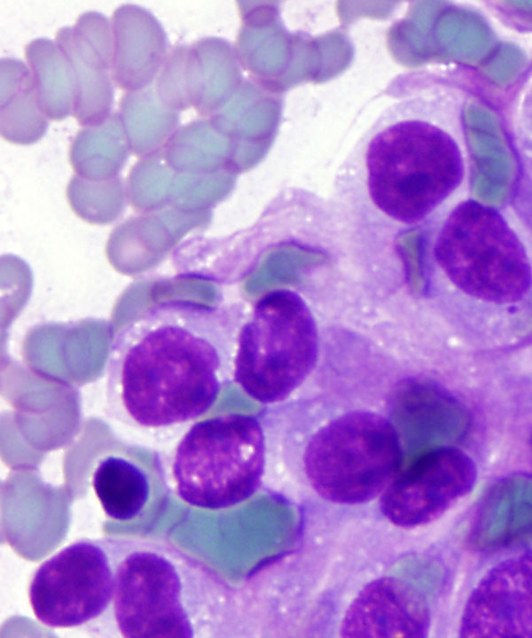
High power: DQ stain
Epithelioid variant GIST
Polygonal cells with round to oval nuclei, and moderate to abundant cytoplasm
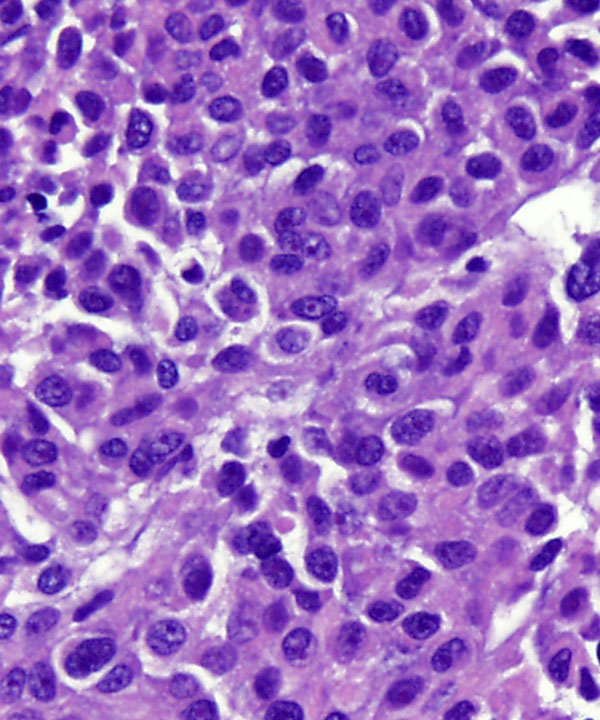
Cell block: H&E
Epithelioid variant GIST
Cell block: IHC for CD117
Epithelioid variant GIST
Features
• Highly cellular smears
• Typically spindled, may be epithelioid or mixed
• Loose or crowded groups
• May have single cells and naked nuclei
• Prominent vascular pattern often present
• Spindle cells
– Uniform with elongated nuclei
– Delicate wispy cytoplasmic processes
– Nuclear palisading may be seen
• Epithelioid cells
– Polygonal with well defined cell borders
– Round/ oval nuclei
– Fine chromatin
– Eosinophilic/ clear cytoplasm
• Cytologic atypia clue to malignancy
• Necrosis and mitosis seen in maligancy
• IHC: Express CD117, DOG1, vimentin, CD34 +/- , usually negative for desmin (D/D smooth muscle tumors), S100 (D/D neural tumors), cytokeratin (D/D spindled carcinoma)
• Mutations in KIT and PDGFRA genes may be present
Dodd LG et al. Am J Clin Pathol. 1998 Apr;109(4):439-43.