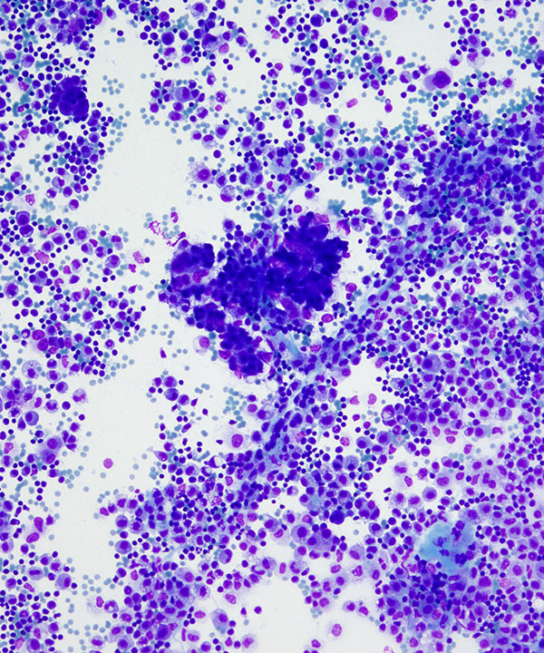
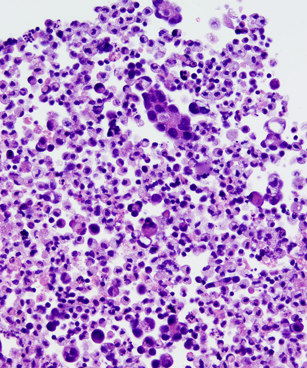
Medium power: DQ stain
3D clusters of crowded malignant cells in a background of histiocytes and inflammatory cells
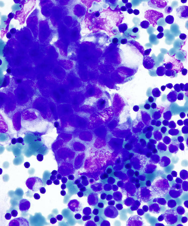
High power: DQ stain
3D cluster of large cells with enlarged nuclei with irregular nuclear membranes and finely vacuolated cytoplasm
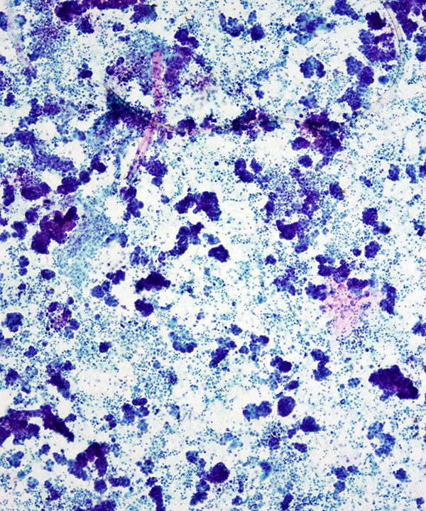
Low power: Pap stain
3D clusters of crowded malignant cells, some showing papillary structures in a background of histiocytes and inflammatory cells
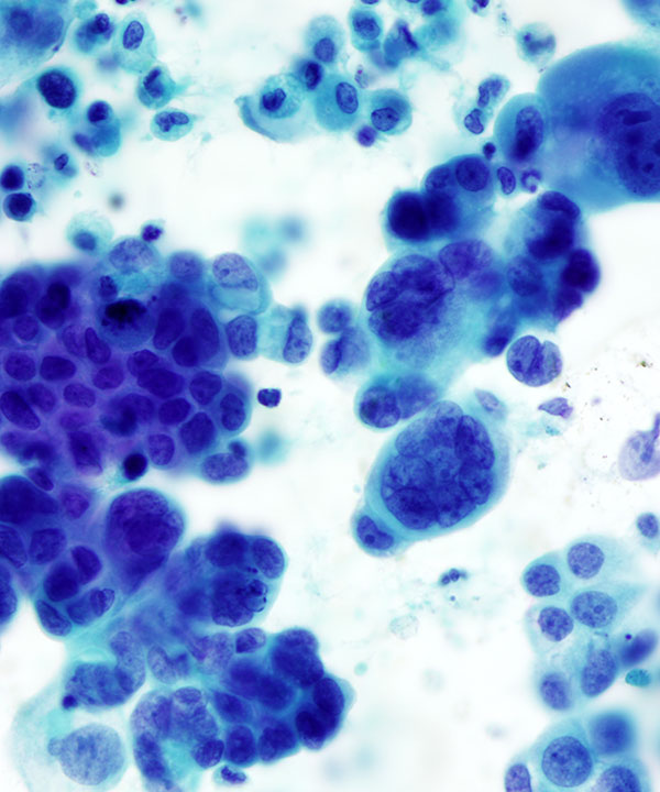
High power: Pap stain
Large malignant cells with pleomorphic enlarged nuclei, irregular nuclear membranes, multinucleation, visible to prominent nucleoli, and finely vacuolated cytoplasm
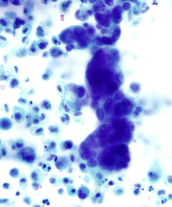
High power: Pap stain
Large malignant cells with high N:C ratios, pleomorphic enlarged nuclei, irregular nuclear membranes, visible to prominent nucleoli, and finely vacuolated cytoplasm
Cell block: H&E stain
Clusters of malignant cells in acinar formations and dispersed singly
Features
• Generally encountered in ascites and peritoneal/pelvic washing specimens.
• Positive cytology modifies Stage I and II to Stage Ic and Iic
• Large and small 3D clusters and single cells
• May demonstrate fibrovascular cores and papillary structures
• Crowded large cells with anisokaryosis
• Scant to moderate finely vacuolated cytoplasm
• May have cytoplasmic mucin vacuoles
• High N:C ratio
• Enlarged hyperchromatic nuclei
• Irregular nuclear membranes
• Coarse chromatin
• Prominent nucleoli
• Mitosis commonly seen
• May have calcifications and psammoma bodies
• IHC: Tumor cells typically express Pax8, ER, while negative for calretinin (D/D mesothelial cells) and CD68 or CD163 (histiocytes). WT-1 is often positive so cannot distinguish from mesothelial cells. Morphologically and immunophenotypically indistinguishable from primary peritoneal serous carcinomas
• Ziselman EM et al. Acta Cytol. 1984 Mar-Apr;28(2):105-10.