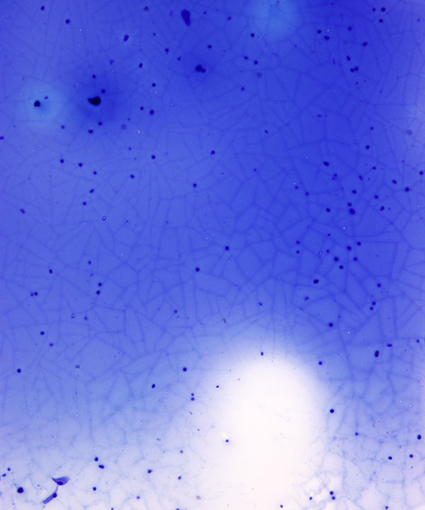
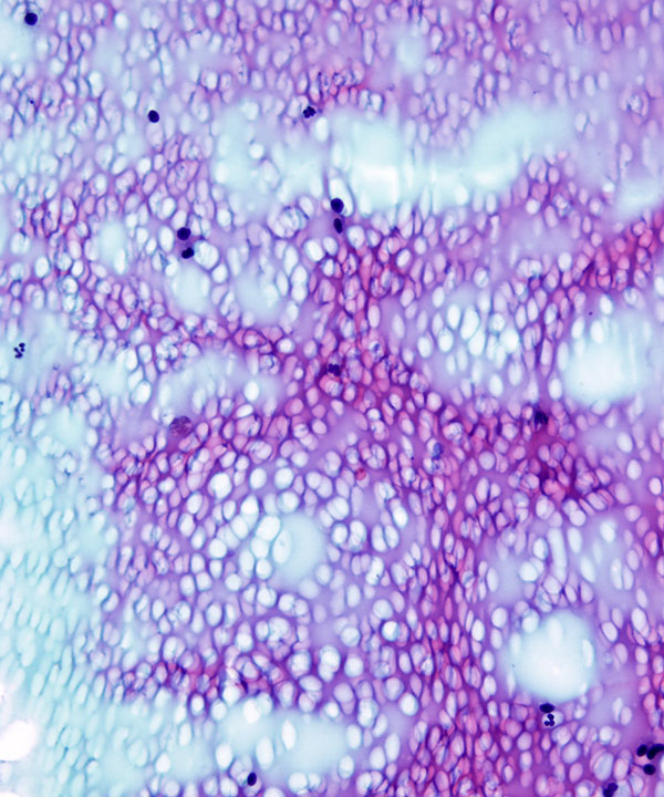
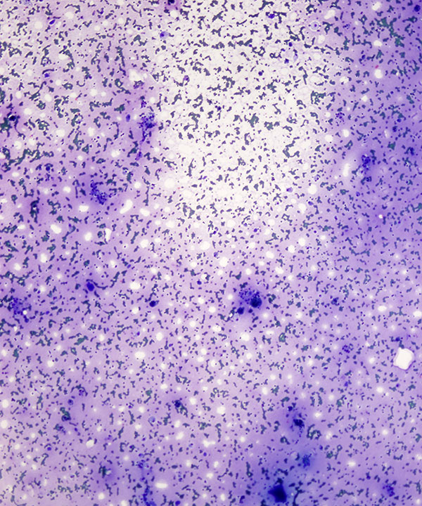
Low power: DQ stain
Watery colloid showing a thin film that cracks on drying
Crazy paving or mosaic appearance
Note scattered histiocytes in the background and lack of follicular epithelial cells
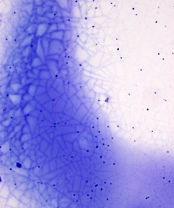
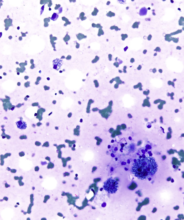
Low power: DQ stain
Watery colloid showing a thin film that cracks on drying
Crazy paving or mosaic appearance
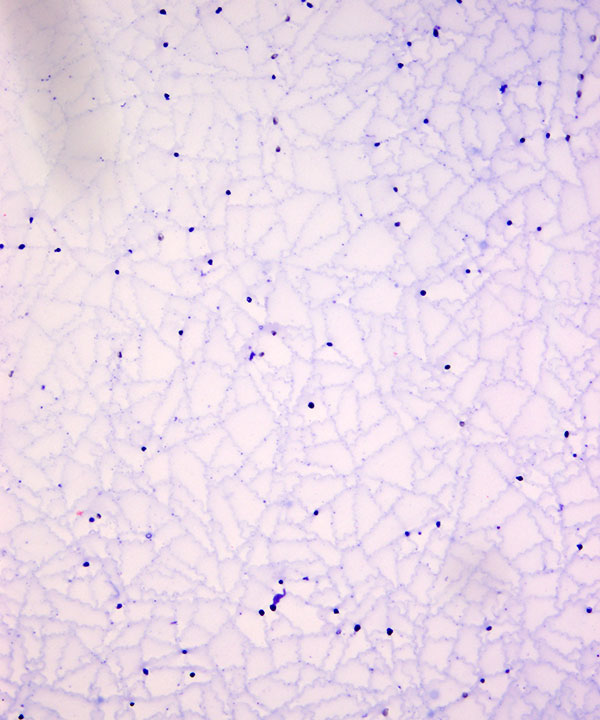
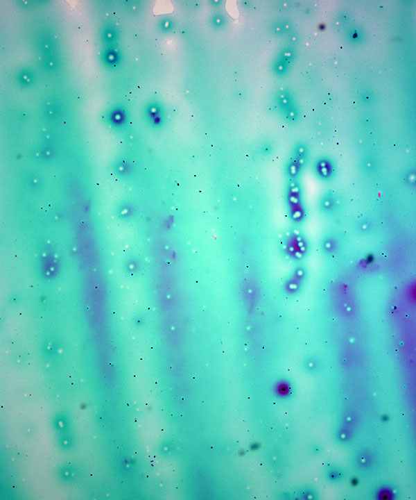
Low power: DQ stain
Watery colloid showing a thin film that cracks on drying
Sometimes the colloid is lost during processing leaving outlines that look like a spider's web or chicken wire
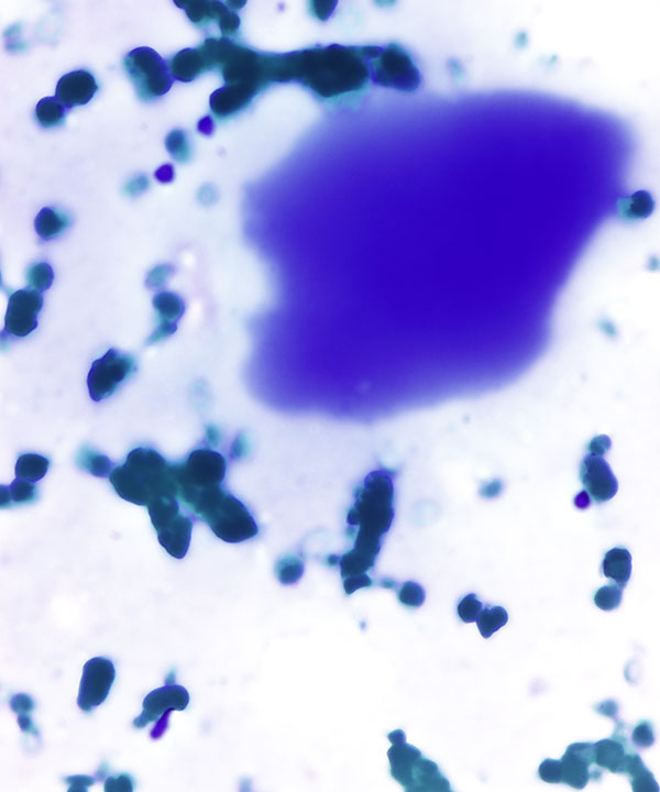
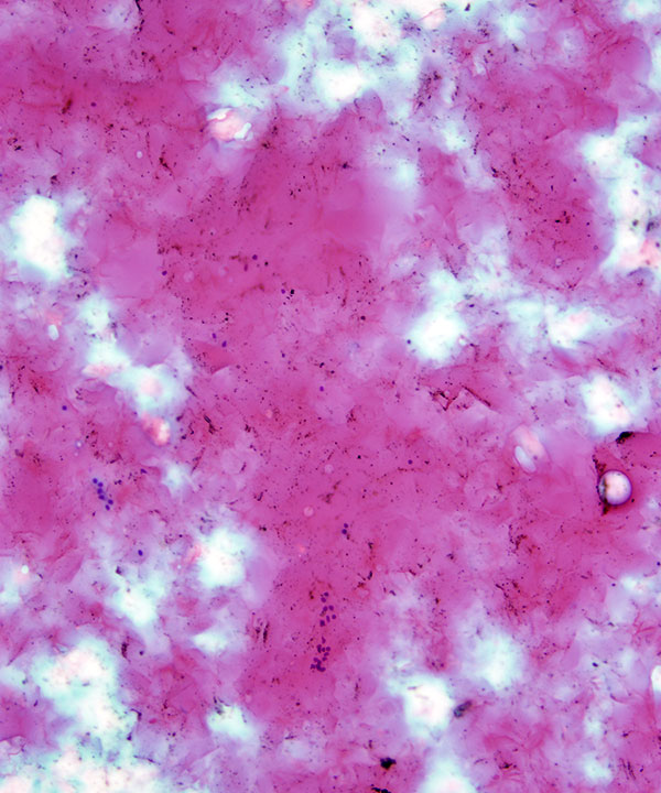
High power: DQ stain
Dense colloid as a homogenous material, often cracks on drying
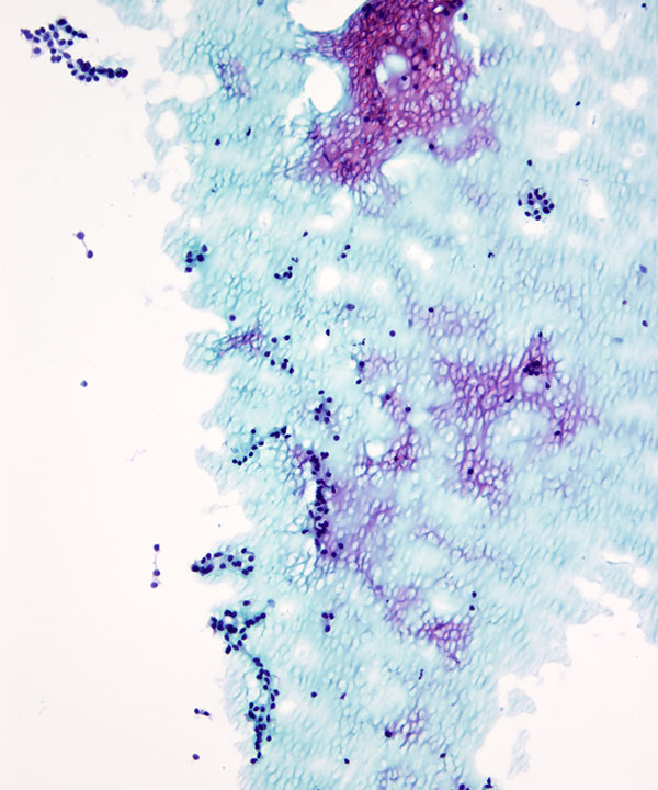
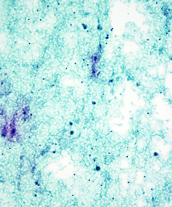
Low power: Pap stain
Watery colloid showing a thin film (blue on the pap stain) that wrinkles and beads up with a mosaic appearance (cellophane-like)
Low power: Pap stain
Watery colloid showing a thin homogenous film (blue on the pap stain)
Abundant colloid favors a colloid nodule
Require cellular criteria of atleast 6 groups of well-visualized follicular cells to make adequacy
Low power: Pap stain
Small clusters and flat sheets of benign follicular cells in a background of watery colloid
Note abundant colloid favors a benign lesion
Low power: Pap stain
Few follicular cells in abundant colloid
Low power: DQ stain
Colloid nodule with cystic degeneration
Scattered hemosiderin-laden macrophages in a background of cyst fluid/colloid
High power: DQ stain
Colloid nodule with cystic degeneration
Hemosiderin-laden macrophages and a few benign follicular epithelial cells
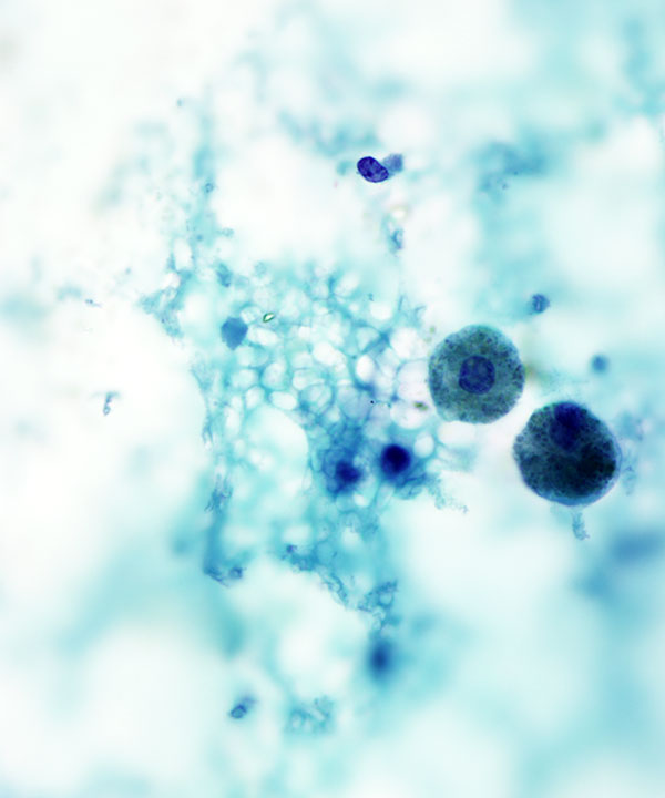
Low power: Pap stain
Colloid nodule with cystic degeneration
Scattered hemosiderin-laden macrophages in a background of cyst fluid/colloid
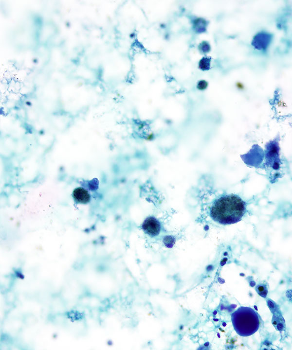
High power: Pap stain
Colloid nodule with cystic degeneration
Hemosiderin-laden macrophages, colloid (dense and watery)
High power: Pap stain
Colloid nodule with cystic degeneration
Hemosiderin-laden macrophages in a background of cyst fluid/colloid
Features
• Sparse to moderately cellular
• Cell to colloid ratio low
• Monolayered flat sheets, 3D balls (spherules), macrofollicles
• May have rare microfollicles
• Uniform small follicular cells
• Scant delicate cytoplasm
• May have paravacuolar blue granules
• May have Hürthle cell change
– Enlarged, eccentric nuclei
– Prominent nucleoli
– Variation in nuclear size
– Nuclear atypia
– Abundant granular cytoplasm
• May have cyst lining cells with reparative change
• Nuclei are small, round, uniform
• No significant atypia or pleomorphism
• Chromatin uniform and granular
• Inconspicuous nucleoli (except Hürthle cells)
• Thin watery colloid
• Dense viscous colloid
• May have cystic changes (histiocytes, multinucleated giant cells)