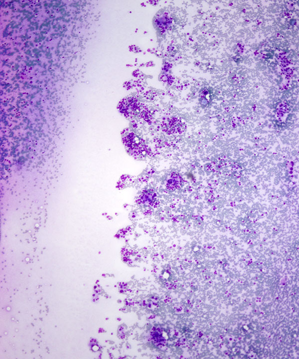
Low power: DQ stain
Cellular smear with single cells and small clusters of oncocytic cells
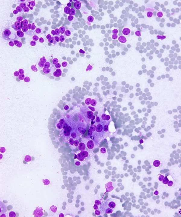
High power: DQ stain
Hürthle cells with round nuclei, frequent binucleation, prominent nucleoli and abundant granular cytoplasm with occasional cytoplasmic vacuoles
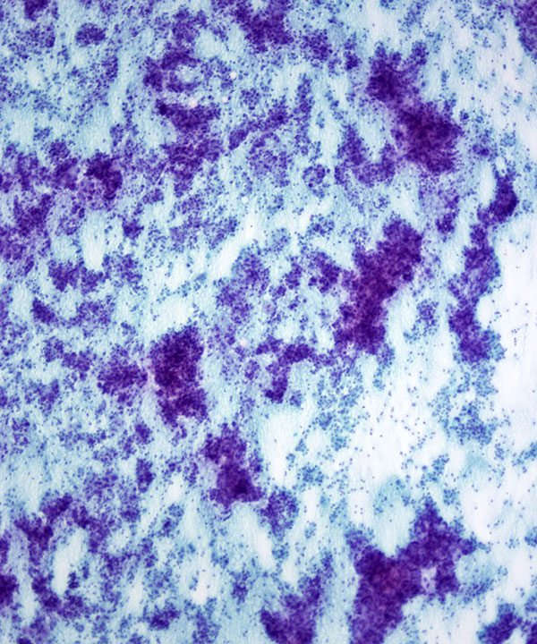
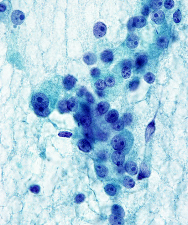
Low power: Pap stain
Cellular smear with numerous single cells and small clusters of oncocytic cells
The cellularity and abundant single cells favor a neoplasm
Note absence of colloid
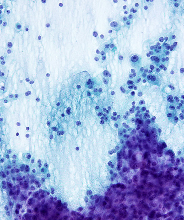
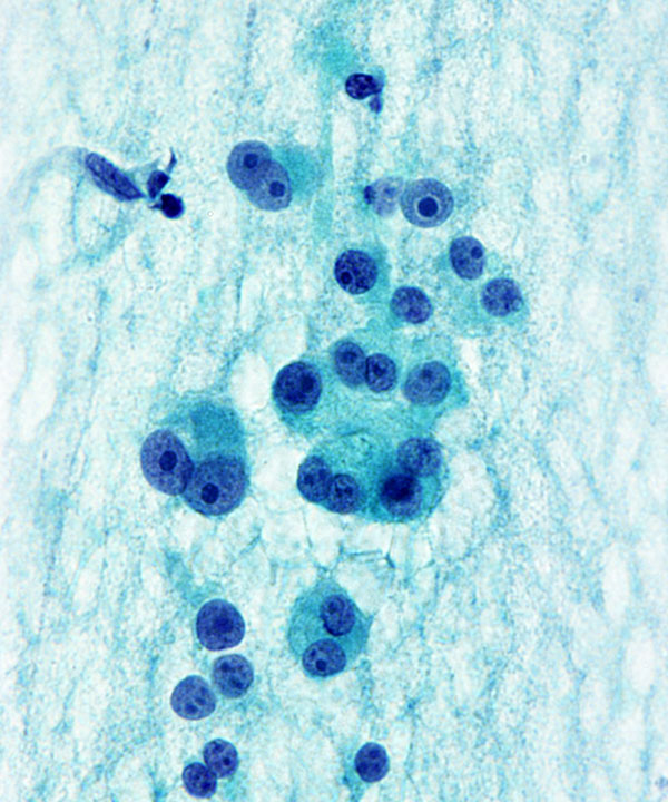
Medium power: Pap stain
Hürthle cells with round eccentrically located nuclei, prominent nucleoli and abundant granular cytoplasm
Note variation in cell sizes with small cells with high N:C ratio and large cells with abundant cytoplasm
Note presence of macronucleoli favor malignancy
High power: Pap stain
Hürthle cells with round nuclei, occasional binucleation, prominent nucleoli and finely granular cytoplasm
High power: DQ stain
Hürthle cells with round nuclei, frequent binucleation, prominent nucleoli and finely granular cytoplasm
Note small cells with high N:C ratio
Note macronucleoli and single cells
Features
• Cellular smears, predominantly Hürthle cells
• Crowded, 3D clusters
• Flat sheets favor benign
• May have papillary architecture
• May have single cells
• Oncocytic cells
• May be bland
• May have atypia or pleomorphism
• Granular cytoplasm
• Intracytoplasmic vacuoles may be present
• High N:C ratio favors malignant
• Nuclei are round/oval,eccentrically located
• Fine to coarse chromatin
• Bi/multinucleation may be present
• Prominent nucleoli present
• Intranuclear cytoplasmic inclusions may be seen
• Scant to absent colloid
• May have cystic change (histiocytes, hemorrhage, giant cells)
• May have naked nuclei (favor malignant)
• Transgressing vessels may be seen
• Similar to follicular neoplasms, cytologic atypia, mitoses and necrosis are not indicators of malignancy. Histologic evidence of invasion or metastasis is required for a diagnosis of Hurthle cell carcinoma.
• Sensitivity of FNA diagnosis of Hürthle cell carcinoma is high but specificity is low, so recommended diagnosis is Follicular neoplasm, Hürthle cell type.
• Express thyroglobulin, may express Galectin-3, S100
Montone KT et al. Arch Pathol Lab Med. 2008 Aug;132(8):1241-50.
Pambuccian SE et al. Acta Cytol. 1997 Jan-Feb;41(1):197-208.
Gonzalez JL et al. Am J Clin Pathol. 1993 Sep;100(3):231-5.
Kini SR et al. Acta Cytol. 1981 Nov-Dec;25(6):647-52.