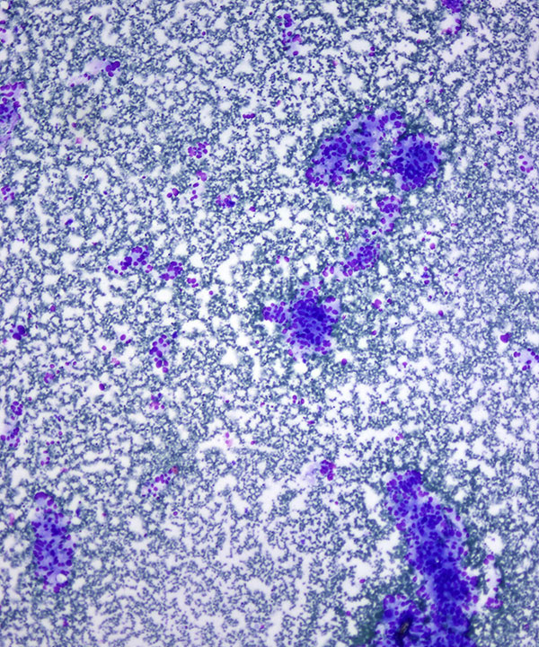
Low power: DQ stain
Loosely cohesive clusters and single cells
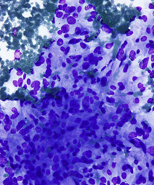
High power: DQ stain
Epithelioid and spindle cells with round to oval nuclei and moderate amounts of cytoplasm
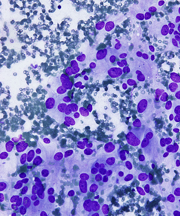
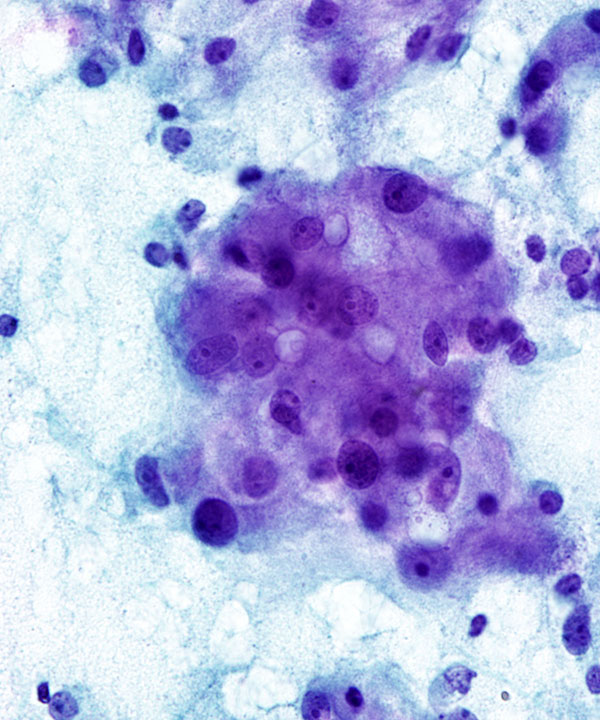
High power: DQ stain
Cells with round to pleomorphic nuclei, binucleation and moderate amounts of cytoplasm
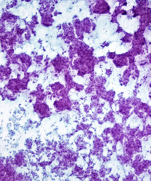
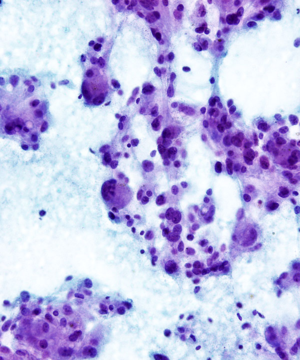
Low power: Pap stain
Cellular smear with loosely cohesive clusters of cells
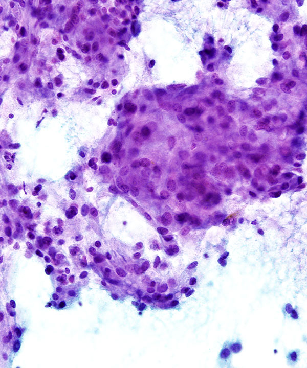
Medium power: Pap stain
Epithelioid and spindle cells with round to oval to pleomorphic nuclei, binucleation and granular cytoplasm
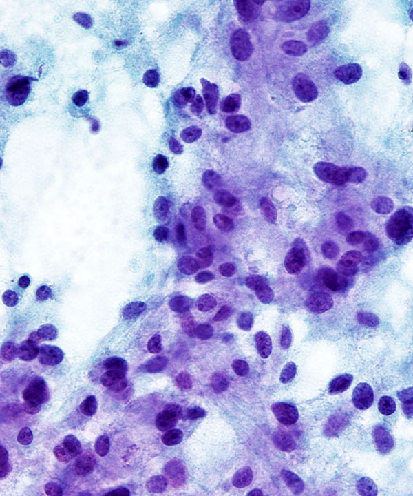
High power: Pap stain
Epithelioid and spindle cells with zellballen appearance, round to oval nuclei and moderate amounts of granular cytoplasm
High power: Pap stain
Epithelioid cells with round to oval nuclei and moderate amounts of granular cytoplasm
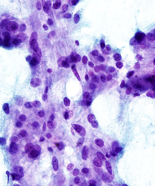
High power: Pap stain
Epithelioid and spindle cells with round to oval nuclei, occasional prominent nucleoli and granular cytoplasm
High power: Pap stain
Cells with granular cytoplasm and occasional intracytoplasmic lipid droplets (D/D adrenocortical tumors)
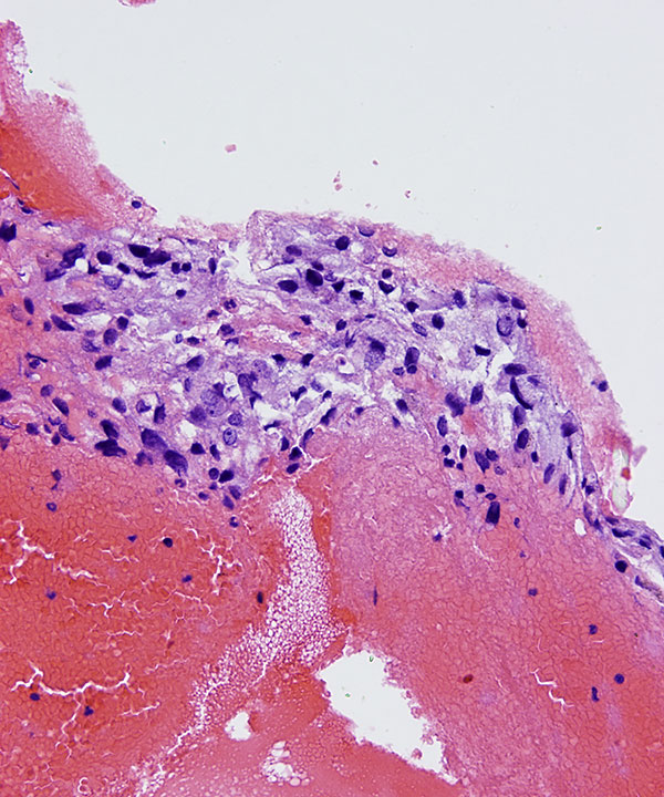
Cell block: H&E stain
Nest of epithelioid and spindle cells with granular cytoplasm
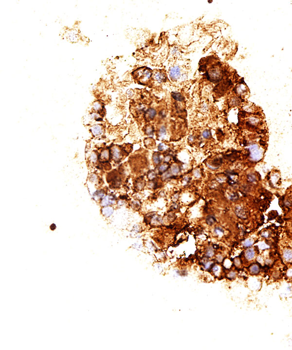
Tumor cells with positive synaptophysin staining (cytoplasmic)
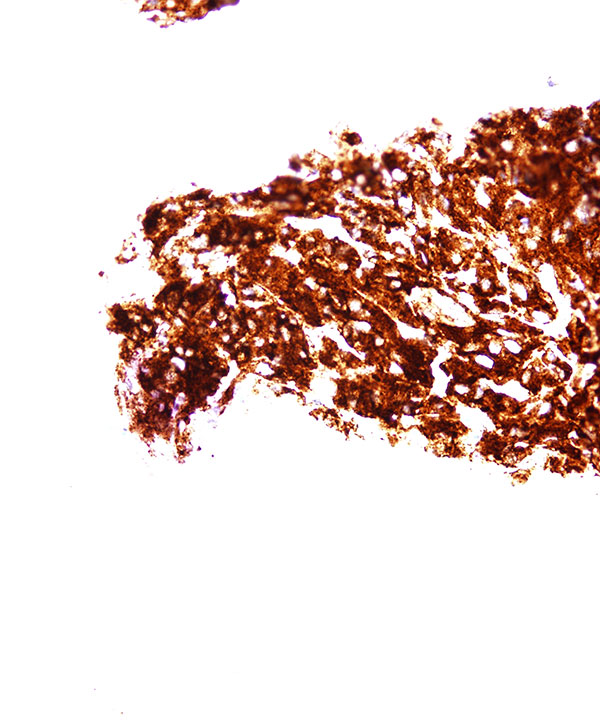
Tumor cells with positive chromogranin staining (cytoplasmic)
Features
• Cellular smears
• Loosely cohesive groups, single cells and rosettes
• May have zellballen (cell ball) appearance
• Anaplasia usually does not correlate with malignancy
• Epithelioid and spindle cells
• Cytoplasm granular with red neurosecretory granules
• May have intracytoplasmic lipid droplets
• May have intracytoplasmic hyaline globules
• Melanin pigment rarely seen
• Nuclei round to oval to pleomorphic
• Binucleation and multinucleation
• May have stippled chromatin (salt and pepper)
• May have prominent nucleoli
• Nuclear atypia is not an indicator of malignancy
• May have mitosis
• May have ganglion cells
• May have spindle/stellate sustentacular cells
• Positive for synaptophysin, chromogranin and CD56.
• Sustentacular cells are usually S100 positive.
• Negative for Inhibin and Melan A, usually negative for cytokeratin.
• RET, VHL, NF1, SDH
• Adrenal cortical adenoma/hyperplasia
• Adrenal cortical carcinoma: inhibin and synaptophysin positive (cytoplasmic), chromogranin negative (cytoplasmic)
• Renal cell carcinoma: pax-8 positive (nuclear), Melan A and inhibin negative (cytoplasmic)
• Metastatic Tumors: more likely to be bilateral, use clinical/radiographic history and site specific immunocytochemical markers if needed
Jimenez-Heffernan JA et al. Acta Cytol. 2006 Jul-Aug;50(4):372-8.
Wagnerova H et al. Bratisl Lek Listy. 2013;114(4):237-40.