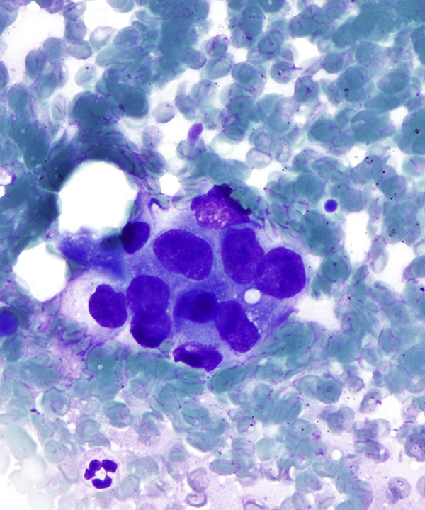
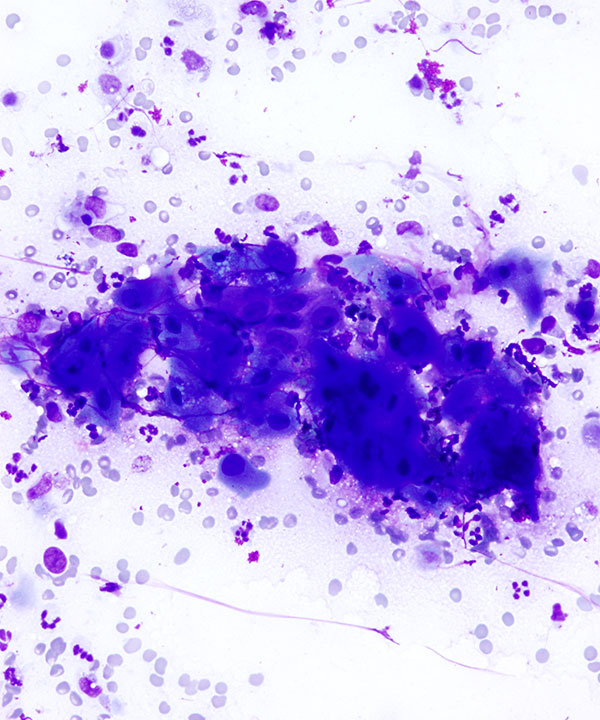
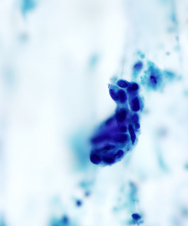
DQ stain: High power
Image showing malignant cells with enlarged irregular nuclei and dense (blue/ purple) cytoplasm
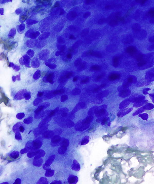
DQ stain: High power
Image showing malignant cells with enlarged irregular nuclei and dense cytoplasm
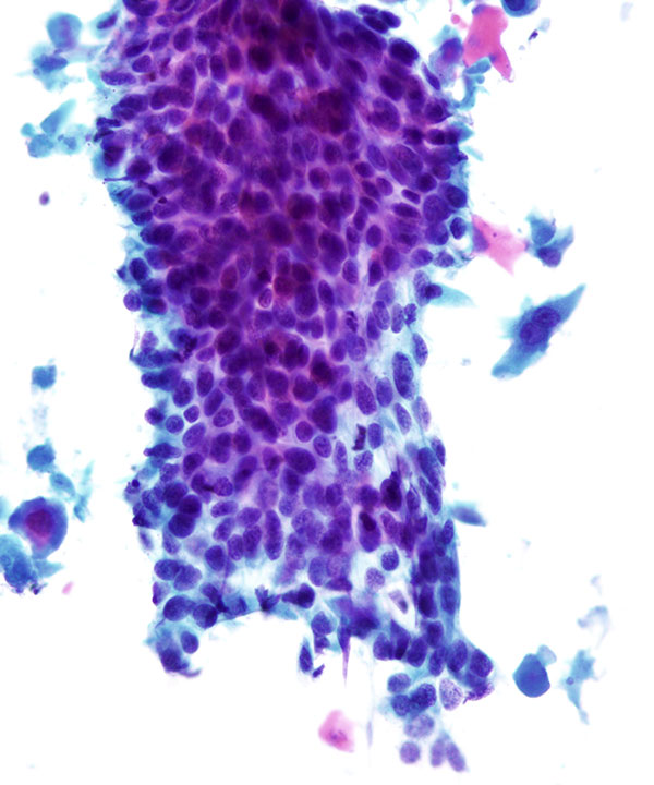
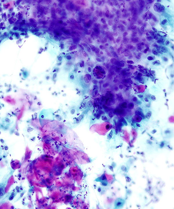
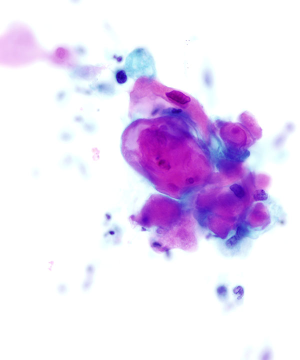
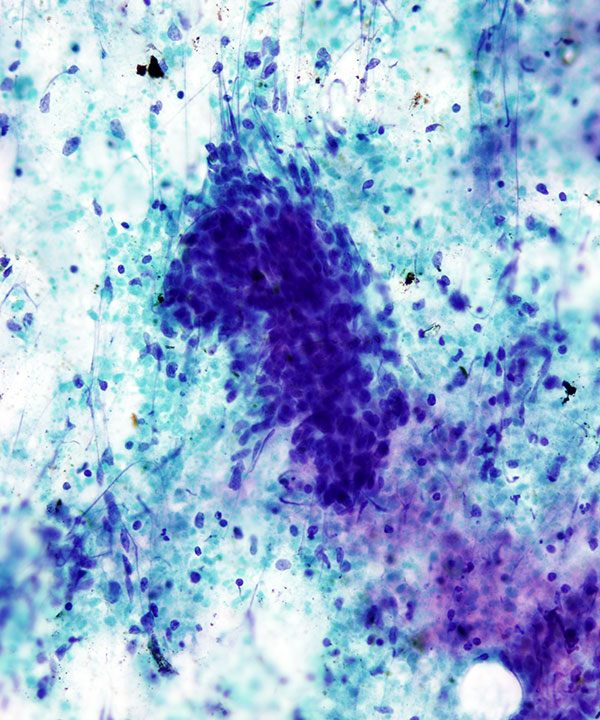
Pap stain: Medium power
Image showing malignant cells in flat sheets with enlarged irregular nuclei, visible to prominent nucleoli and dense cytoplasm.
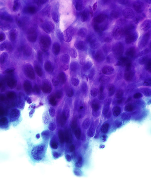
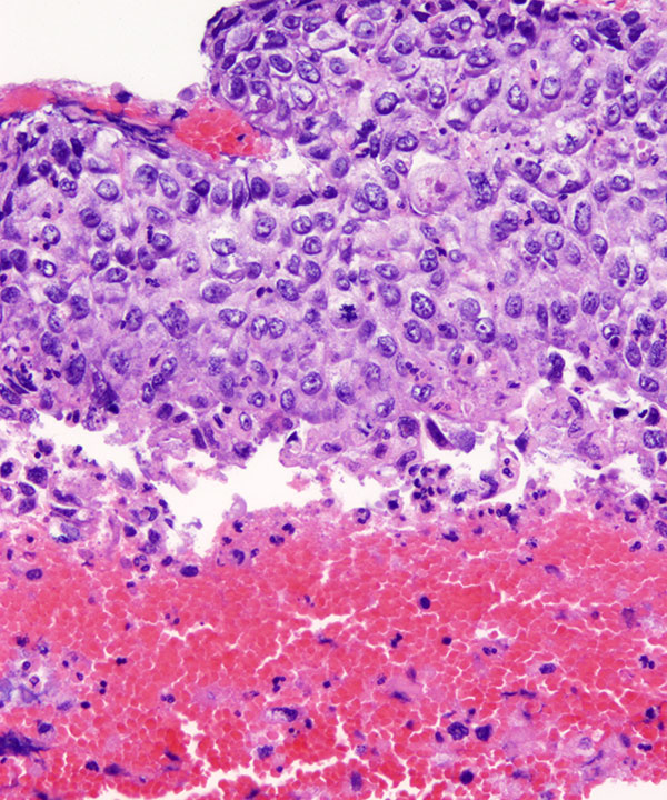
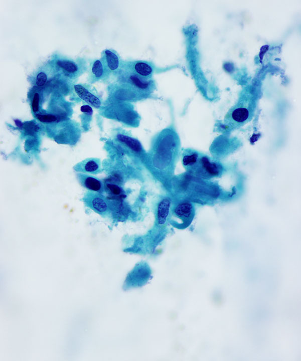
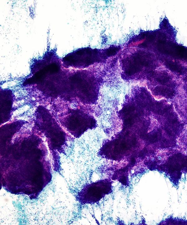
Pap stain: High power
Image showing malignant cells in sheets with enlarged irregular nuclei, visible to prominent nucleoli and dense cytoplasm. Note distinct cell borders.
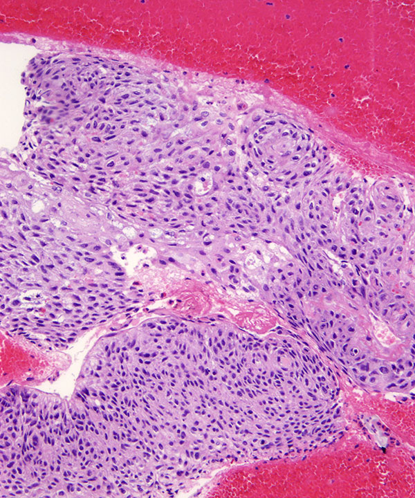
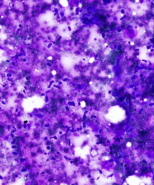
H&E stained cell block showing nests of malignant cells.
H&E stained cell block showing poorly differentiated squamous carcinoma with nuclear pleomorphism, dyskeratitic cells and mitotic figures.
Variants
Keratinizing squamous cell carcinoma: Medium power DQ stain
Keratinized squamous cells with dense cytoplasm
Variants
Keratinizing squamous cell carcinoma: Medium power Pap stain
Keratinized squamous cells
Variants
Keratinizing squamous cell carcinoma: High power Pap stain
Keratinized debris and carcinoma cells

Variants
Keratinizing squamous cell carcinoma: Cell block H&E stain
Keratinized squamous cells with keratin pearls
Variants
Spindle cell variant: Medium power DQ stain
Spindle cells in a background of necrosis
Variants
Spindle cell variant: High power Pap stain
Spindle cells in a small cluster
Variants
Spindle cell variant: High power Pap stain
Spindle cells with dense cytoplasm and cytoplasmic processes (tails)
Variants
Basaloid variant: Medium power Pap stain
Basaloid cells in a background of necrosis
Variants
Basaloid variant: Low power Pap stain
Basaloid cells in aggregates and nests in a background of necrosis
Features
• Flat sheets
• Groups and single cells
• Dense cytoplasm
• Distinct cell borders
• Enlarged, irregular nuclei
• Often pleomorphic
• Visible to prominent nucleoli
• Coarse chromatin
• Usually necrotic
• Squamous pearls, dyskeratotic cells, bizarre cells, necrotic debris, granulomas
• Keratinizing, non-keratinizing, basaloid, spindle cell
• Express cytokeratin, CK5/6, high molecular weight keratin, p63, p40; usually negative for TTF-1, CK7, CK20
Zusman-Harach SB et al. J Clin Pathol. 1991 Dec;44(12):997-1002.
Kamiya M et al. Acta Cytol. 1995 Jan-Feb;39(1):61-8.