
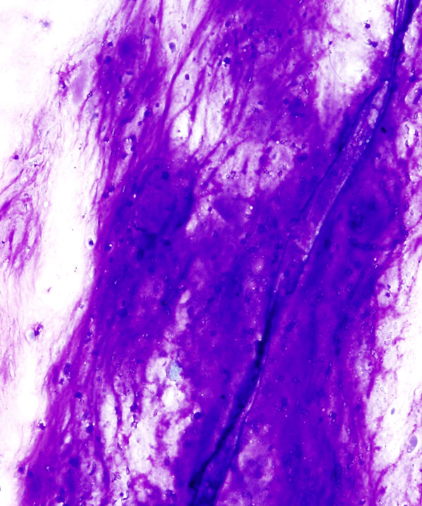
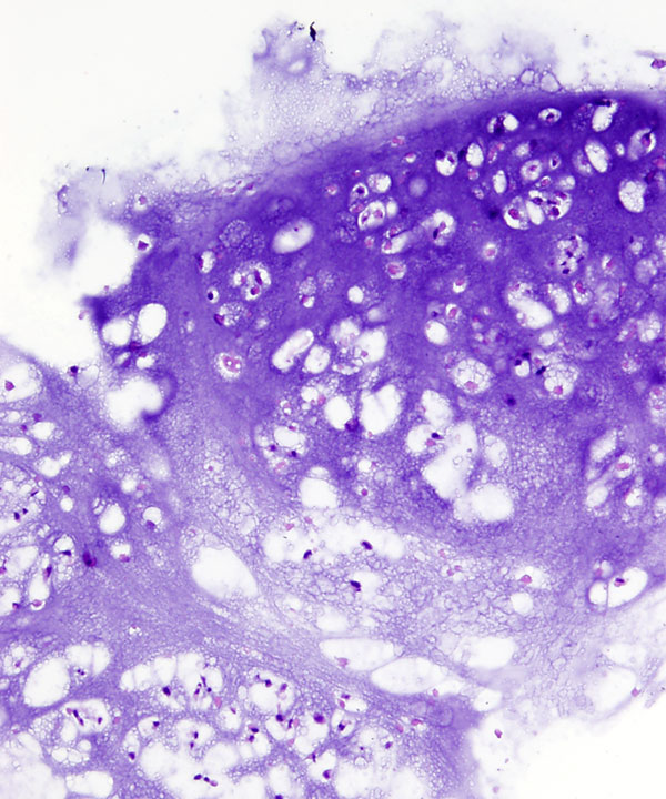
DQ stain: Low power
Metachromatic cartilaginous matrix
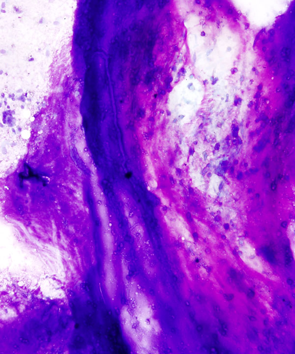
DQ stain: Low power
Singly dispersed small uniform cells in a background of cartilaginous matrix (metachromatic on DQ stain)
DQ stain: Low power
Singly dispersed chondrocytes in a background of cartilaginous matrix
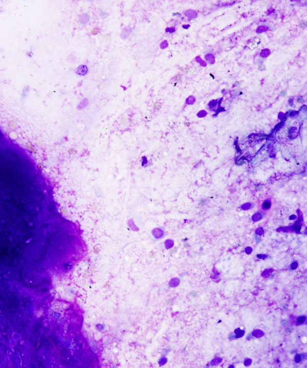
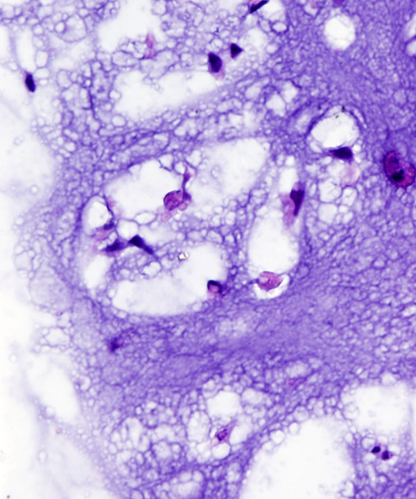
DQ stain: Medium power
Singly dispersed small uniform cells in a background of cartilaginous matrix (metachromatic on DQ stain)
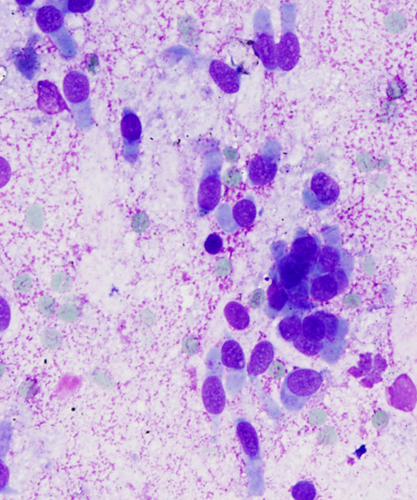
DQ stain: High power
Small uniform cells with round to oval nuclei and abundant vacuolated to granular cytoplasm
Pap stain: Low power
Chondrocytes in lacunar spaces embedded in cartilaginous matrix
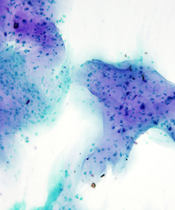
Pap stain: Low power
Loosely cohesive and singly dispersed cells in a background of cartilaginous matrix
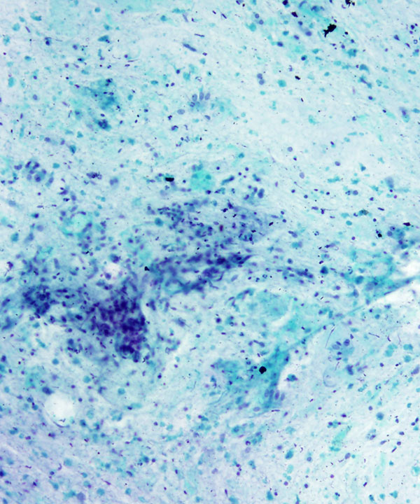
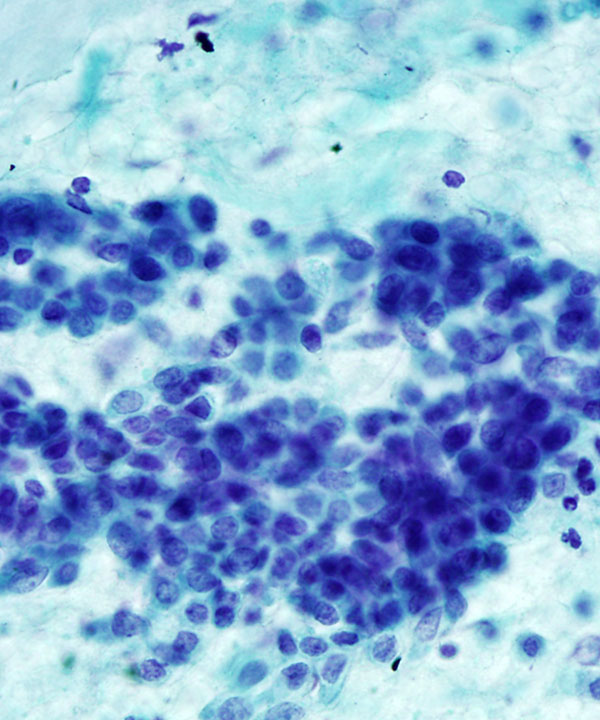
Pap stain: Low power
Loosely cohesive and singly dispersed small uniform cells in a background of cartilaginous matrix
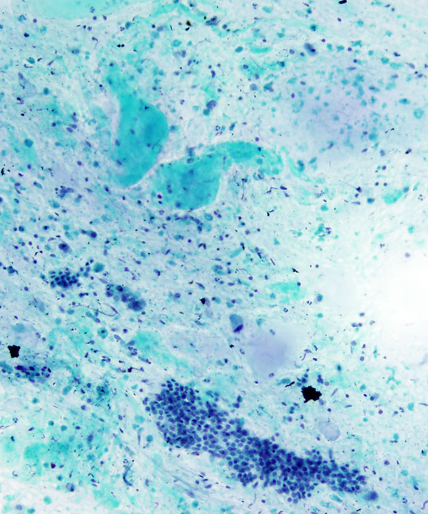
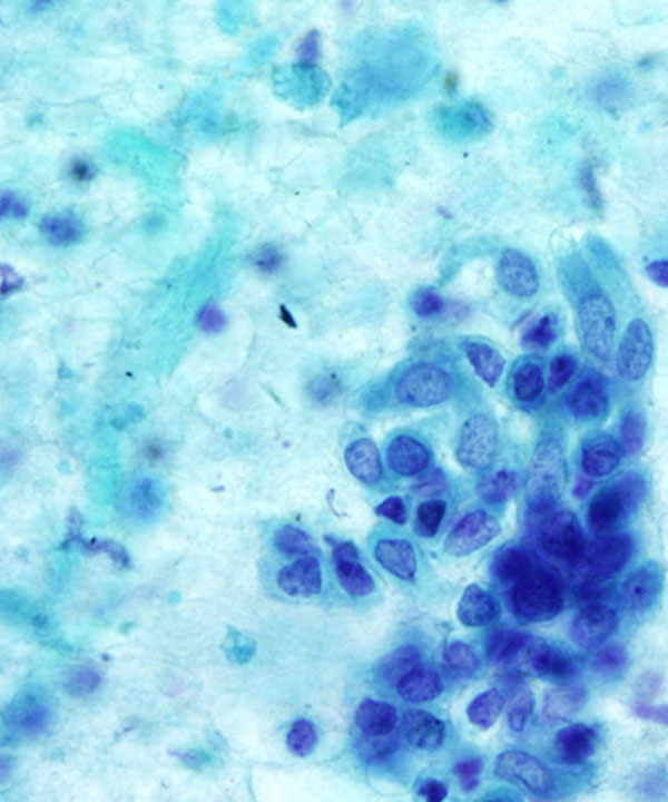
Pap stain: Medium power
Small uniform cells with round to oval nuclei, fine to coarse chromatin, visible nucleoli and vacuolated cytoplasm
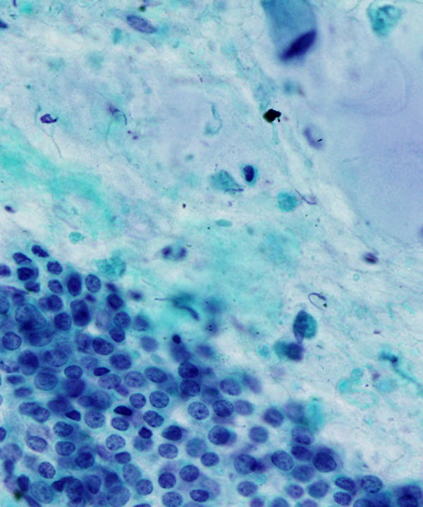
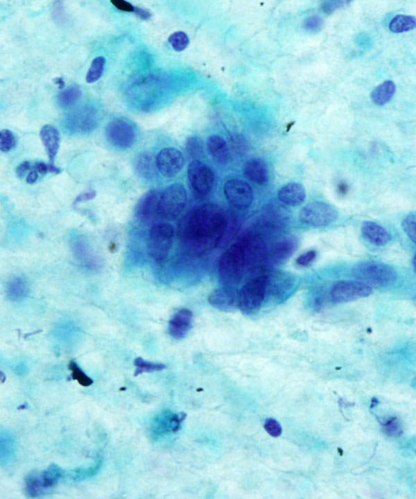
Pap stain: Medium power
Loosely cohesive clusters of small uniform cells with round to oval nuclei, fine to coarse chromatin, visible nucleoli and vacuolated cytoplasm. Nuclear atypia present.
Pap stain: Medium power
Loosely cohesive clusters of chondrocytes with nuclear atypia. Note the distinct cell borders.
Pap stain: Medium power
Loosely cohesive clusters of chondrocytes with nuclear atypia in lacunar spaces. Note the distinct cell borders.
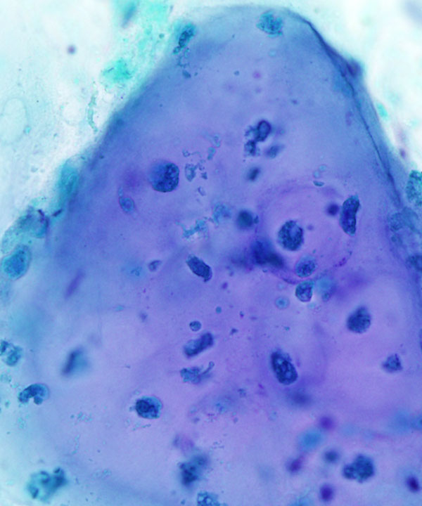
Pap stain: Medium power
Chondrocytes embedded in cartilaginous fragment
Cell block: H&E stain
Chondrocytes in lacunar spaces
Cell block: H&E stain
Malignant chondrocytes with nuclear atypia in lacunar spaces
Features
• Hyaline cartilage malignant tumor
• Occurs in bone and soft tissue
• Presents with pain and tenderness
• Often intermediate grade with indolent course
• Pleomorphism may be seen with high grade or dedifferentiated tumors
• Variable cellularity
• Chondrocytes embedded in metachromatic chondroid matrix
• Loose clusters or singly dispersed
• Oval to polygonal chondrocytes
• Abundant vacuolated to granular cytoplasm
• Distinct cell boundaries
• Nuclei round to oval, uniform
• Nuclear atypia in higher grade tumors
• May have binucleation/ multinucleation
• Fine to coarse chromatin
• Prominent nucleoli
• Cartilaginous matrix
• May have osteoclastic giant cells
• Usually calcification/ossification absent
• Olszewski W. et al. Acta Cytol. 1983 May-Jun;27(3):345-9.
• Abdul-Karim FW et al. Acta Cytol. 1993 Sep-Oct;37(5):655-60.