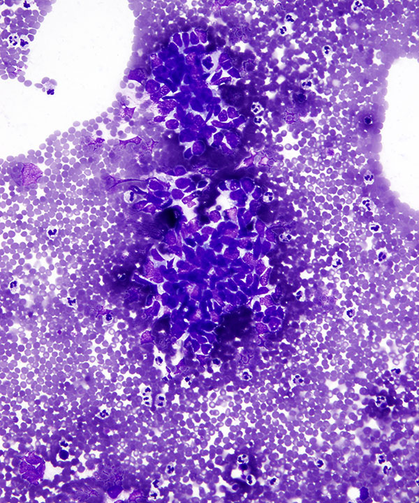
Low power: DQ stain
Cluster of malignant cells with high N:C ratios, nuclear molding and scant cytoplasm
Diagnosis: Metastatic small cell carcinoma
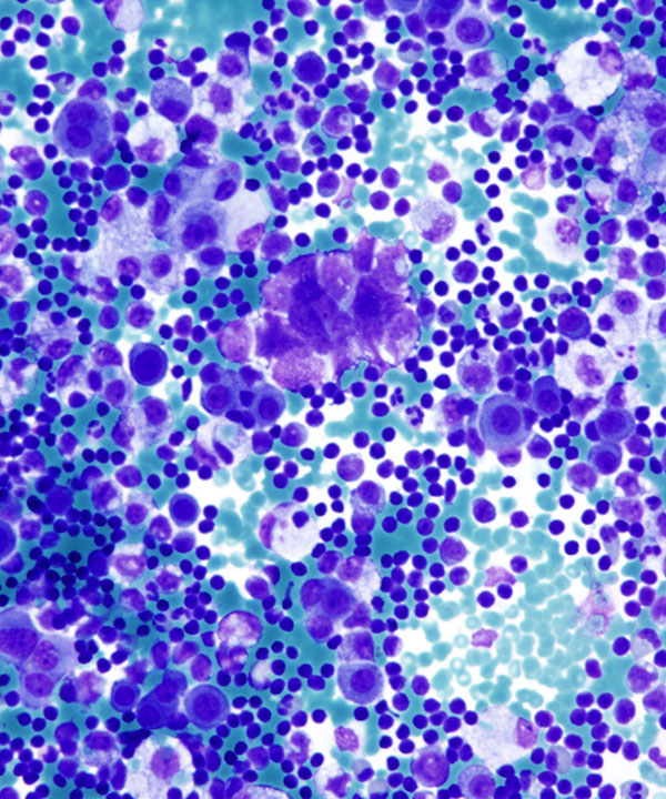
Medium power: DQ stain
Cluster of malignant cells with high N:C ratios, nuclear molding and scant cytoplasm in a background of lymphocytes and histiocytes
Diagnosis: Metastatic small cell carcinoma
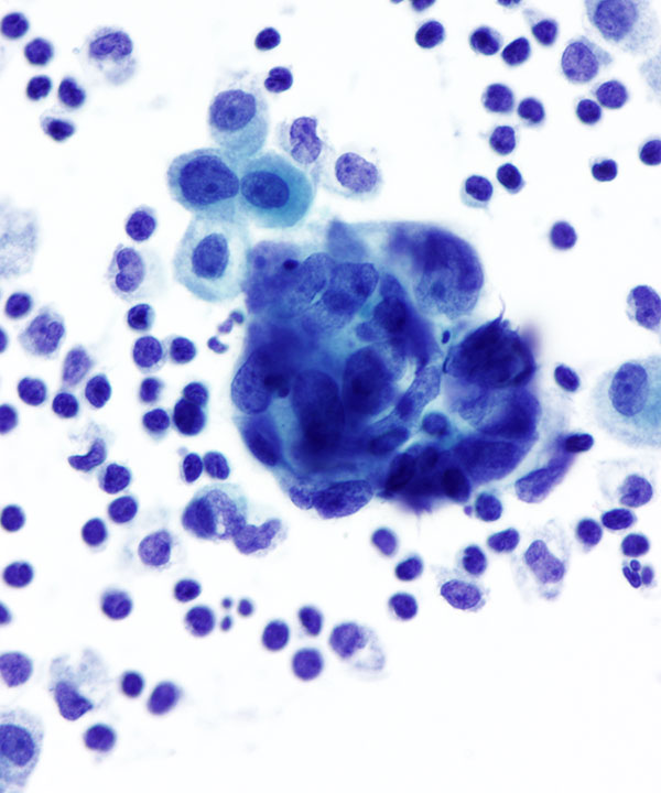
Medium power: Pap stain
Cluster of malignant cells with high N:C ratios, nuclear molding and scant cytoplasm in a background of lymphocytes and histiocytes
Diagnosis: Metastatic small cell carcinoma
Small cell carcinoma can present as small tumor cells that can blend in with the background and be missed
Features
• Most common cause of malignant effusions with adenocarcinomas being the most frequent.
• Foreign population of cells that stand out from mesothelial cells and histiocytes
• Exception:
- cells that mimic native cells
- all tumor cells so they all look alike
• Carcinomas typically form cohesive clusters and ball up to form spheres with smooth community borders ; however some tumors shed as single cells (lobular breast carcinoma, gastric signet ring cells). May or may not have malignant features (enlarged irregular nuclei with pleomorphism) but usually have high N:C ratios and coarse chromatin.
• High N:C ratios
• Single cells, small clusters
• Cells only slightly larger than lymphocytes
• Scant cytoplasm
• Cells angulated and wrap around (molding) themselves
• Nuclei enlarged, hyperchromatic
• Nuclear molding
• Finely granular chromatin
• Inconspicuous to absent nucleoli
• IHC: Tumor cells typically express TTF-1, CD56, synaptophysin, chromogranin, while negative for calretinin (D/D mesothelial cells), CD68 or CD163 (histiocytes), CK20 (D/D Merkel cell) and CD45 (D/D lymphoma)