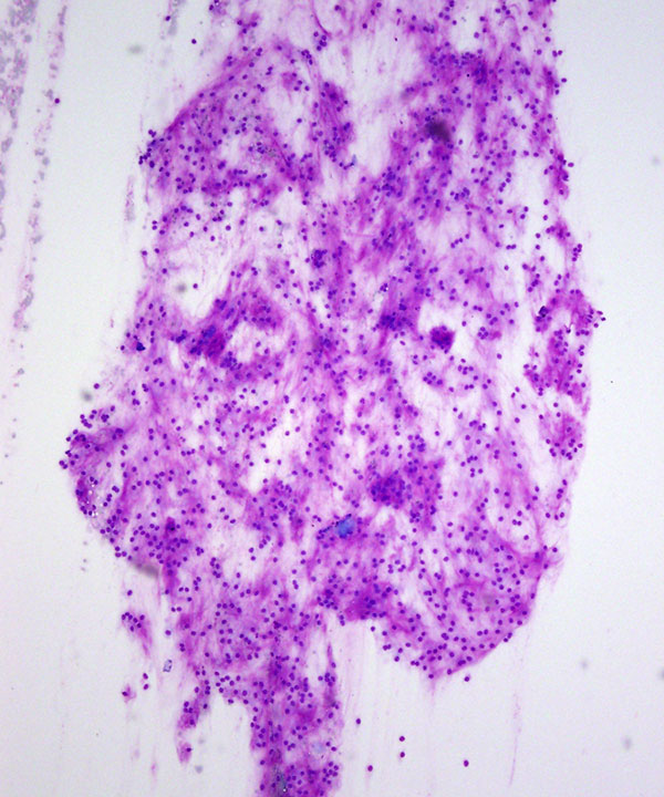
Low power: DQ stain
Moderately cellular smear, fragments of fibrillary stroma
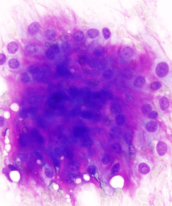
High power: DQ stain
Highly cellular, condensed magenta colored stroma
High power: DQ stain
Relatively less cellular, fibrillary magenta colored stroma
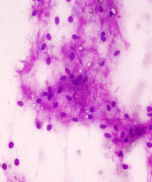
High power: DQ stain
Fibrillary, magenta colored stroma; note the single cell filing pattern
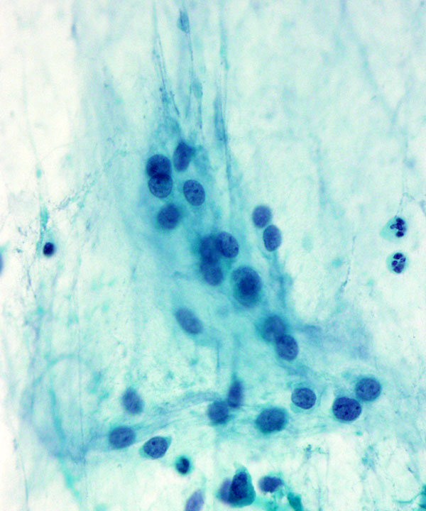
High power: Pap stain
Less prominent stroma; single cell filing pattern of the cells
Note the uniformity of nuclei and single small nucleolus
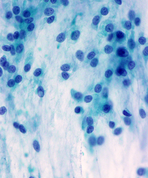
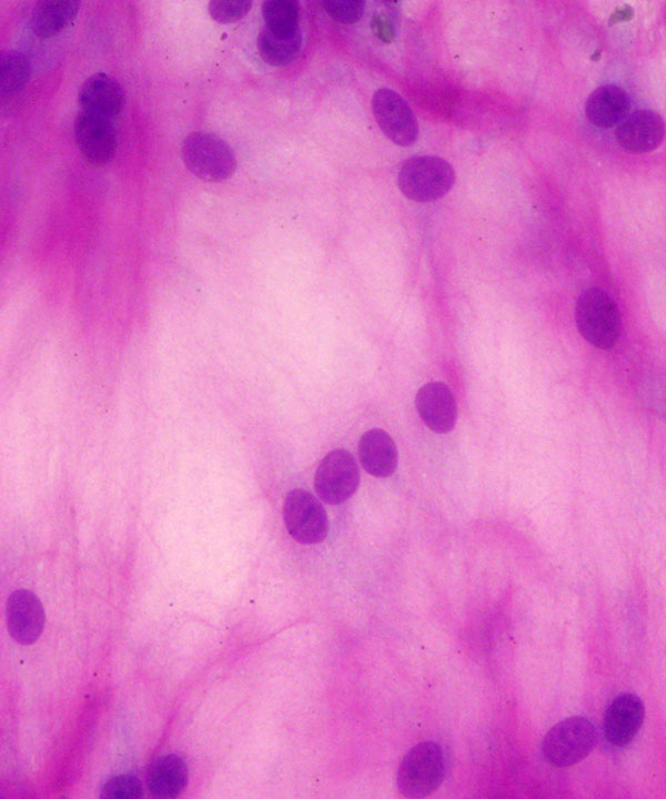
High power: Pap stain
Indistinct stroma; note the uniform nuclei, small nucleolus, nuclear grooves and pseudoinclusion
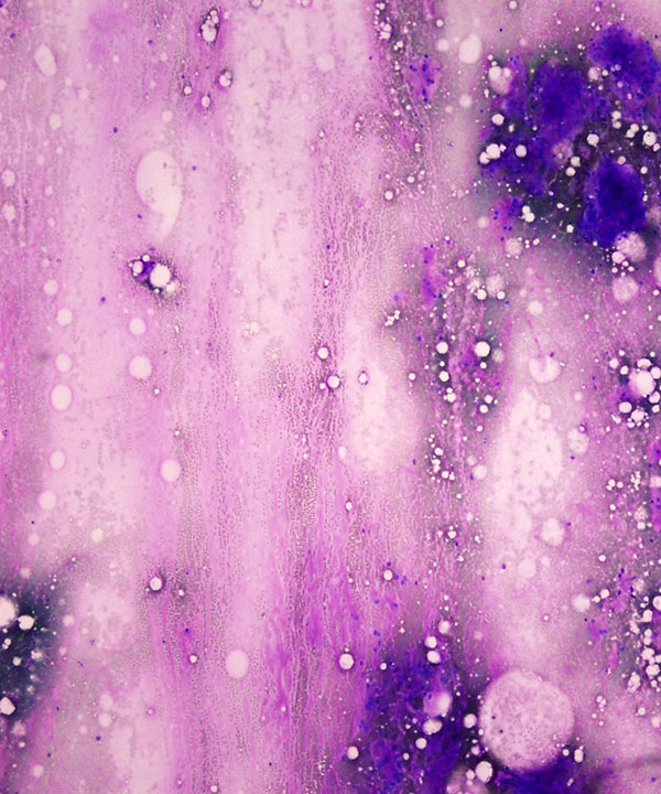
Low power: DQ stain
Thick fragments of magenta colored stroma (upper half)
Thin, streaming, myxoid stroma (lower half)
Bubble artifact (NOT to be confused with adipocytes)
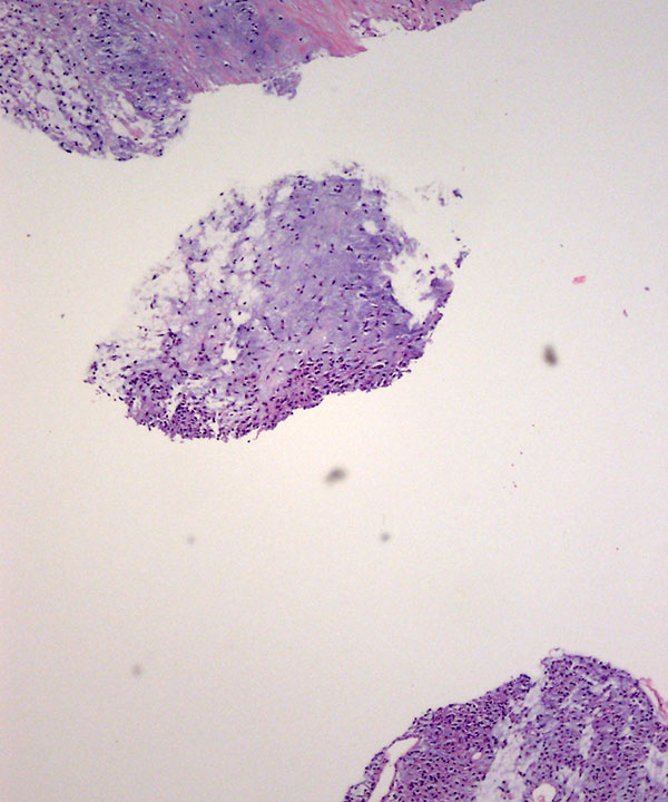
Low power: Cell block H&E stain
Variability in the cellularity: Hypocellular fragment (left), variably cellular fragment (middle); hypercellular fragment (right)
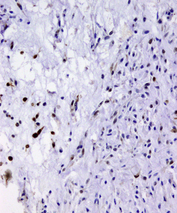
High power: Cell block, S-100 stain
Focal nuclear and cytoplasmic staining, note the difference in the cellularity between upper right and lower left halves of this fragment
Features
• Moderate cellularity of the smear
• Cohesive fragments of fibrillary stroma
• Variable cellularity of stromal fragments
• Small cords of cells (single cell filing pattern)
• Ovoid, bland spindle cells
• Minimal cytoplasm
• Long wispy cytoplasmic extensions which intertwine
• Distinct cell borders
• Uniform nuclei (no pleomorphism)
• Eccentric location of nuclei
• Ovoid shape
• Nuclear grooves and pseudo-inclusions
• Single, indistinct nucleolus
• Condensed, magenta colored fibrillary matrix (seen on DQ stain)
• Less apparent matrix on Pap stain
• Background of thin myxoid material
• Bubble artifact during smearing due to thickness of matrix
Thool A et al . Acta Cytol. 2001 Jan-Feb;45(1):86-8.