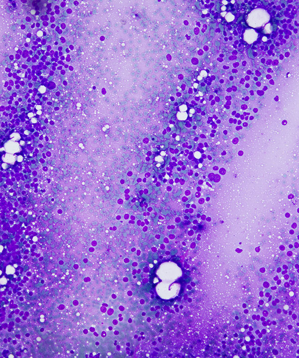
Low power: DQ stain
Cellular smear with numerous single cells
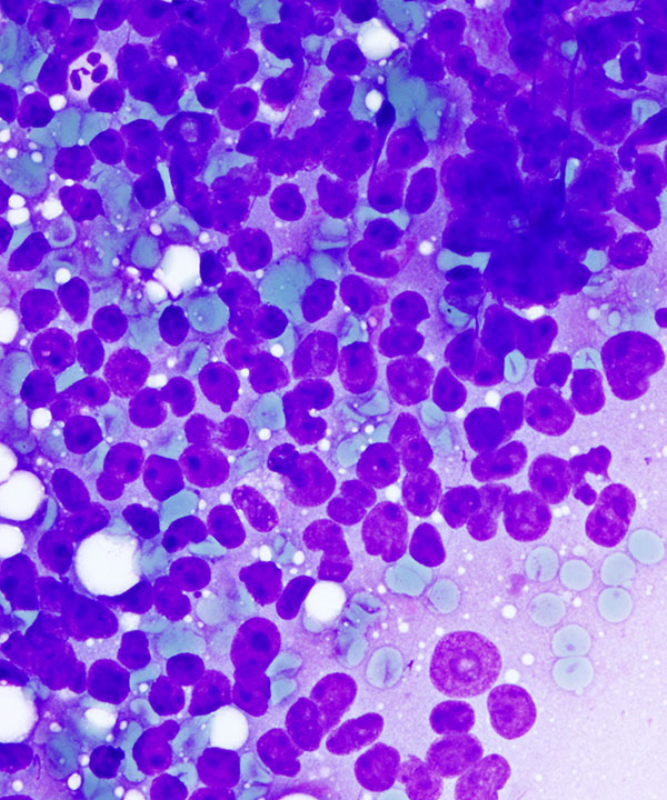
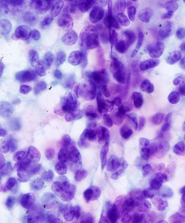
High power: DQ stain
Cellular smear with numerous single cells, enlarged irregular nuclei and foamy cytoplasm
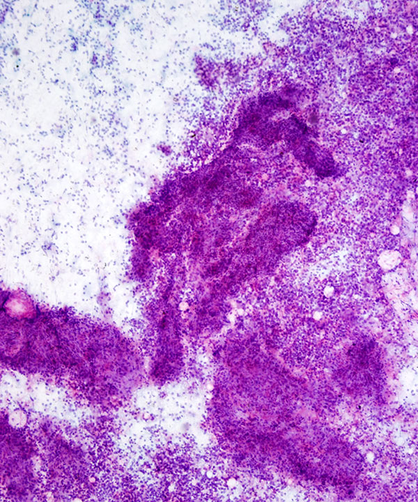
Low power: Pap stain
Cellular smear with cohesive clusters and numerous single cells
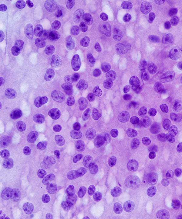
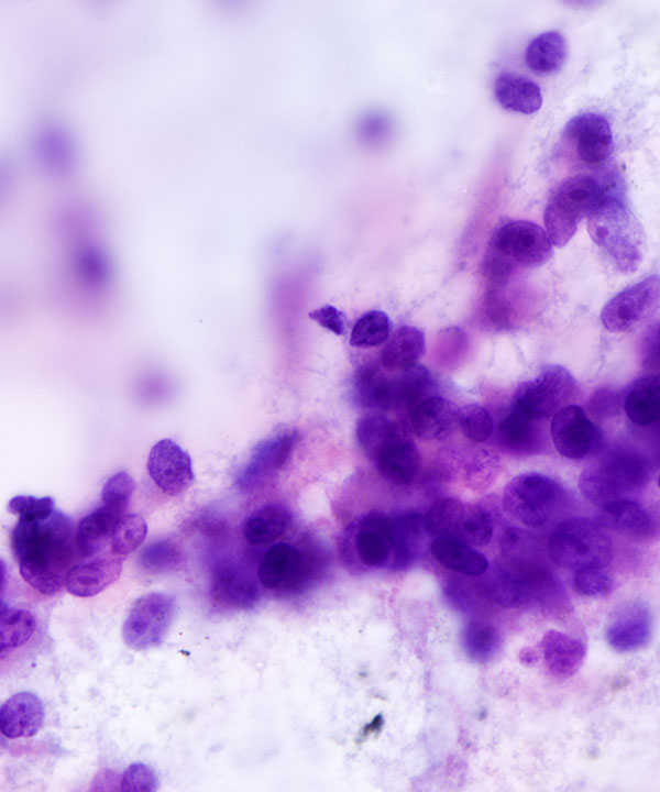
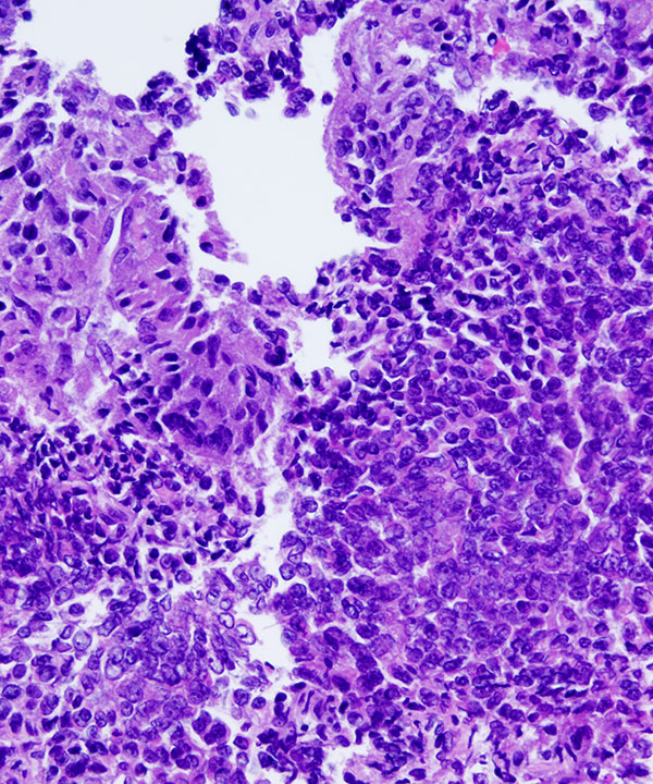
High power: Pap stain
Numerous single cells with enlarged irregular nuclei and prominent nucleoli
High power: Pap stain
Numerous single cells with enlarged irregular nuclei and prominent nucleoli
Note endothelial cells of vessels traversing cell clusters
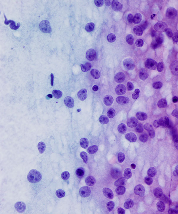
High power: Pap stain
Numerous single cells with enlarged irregular nuclei and prominent nucleoli
Note mostly preserved cytoplasm
High power: Pap stain
Numerous single cells with enlarged irregular nuclei, coarse chromatin, and prominent nucleoli
Note mitosis (upper right)
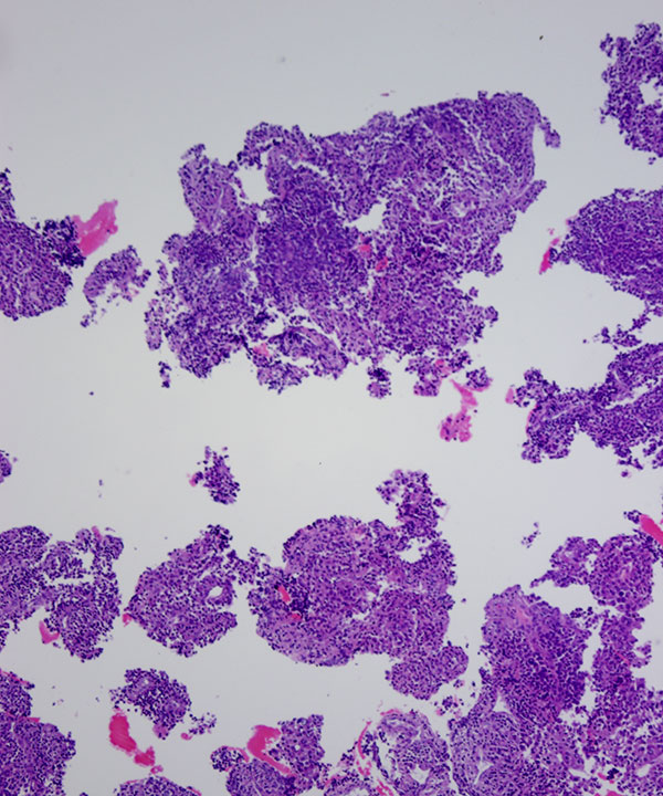
Cell block: H&E stain
Cells are typically positive for inhibin (cytoplasmic), Melan A (cytoplasmic), and calretinin (cytoplasmic and nuclear)
Cell block: H&E stain
Cells are typically positive for inhibin (cytoplasmic), Melan A (cytoplasmic), and calretinin (cytoplasmic and nuclear)
Features
• Diffuse growth pattern
• Cellular smear with cohesive clusters and single intact cells
• Capillaries traversing and wrapping cell clusters
• Bland low N:C ratio cells to anaplastic malignant looking cells
• Epithelioid or spindled
• May have multinucleated bizarre cells
• Cytoplasm vacuolated but usually preserved
• May have granular cytoplasm
• Nuclei may be enlarged, hyperchromatic, irregular, pleomorphic
• Coarse chromatin
• Prominent nucleoli
• May have intranuclear cytoplasmic inclusions
• Naked nuclei less common
• Mitosis common, including atypical mitoses
• Usually necrosis present
• Foamy background less common
• Positive stains: inhibin (cytoplasmic), Melan A (cytoplasmic), calretinin (cytoplasmic and nuclear), synaptophysin +/- (cytoplasmic)
• Negative stains: chromogranin (cytoplasmic), pax-8 (nuclear)
• Adrenal cortical adenoma/hyperplasia
• Pheochromocytoma: chromogranin positive (cytoplasmic), Melan A and inhibin negative (cytoplasmic)
• Renal cell carcinoma: pax-8 positive (nuclear), Melan A and inhibin negative (cytoplasmic)
• Metastatic Tumors: more likely to be bilateral, use clinical/radiographic history and site specific immunocytochemical markers if needed
Saboorian MH et al. Acta Cytol. 1995 Sep-Oct;39(5):843-51.
Tirabassi et. al. J Endocrinol Invest. 2012 Jun;35(6):590-4.
Wagnerova H et al. Bratisl Lek Listy. 2013;114(4):237-40.