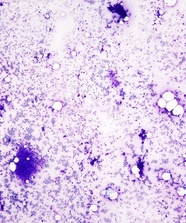
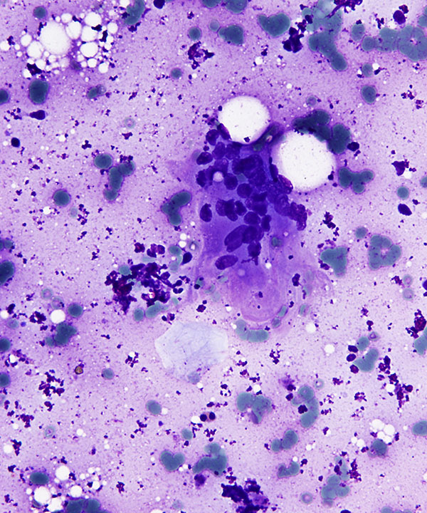
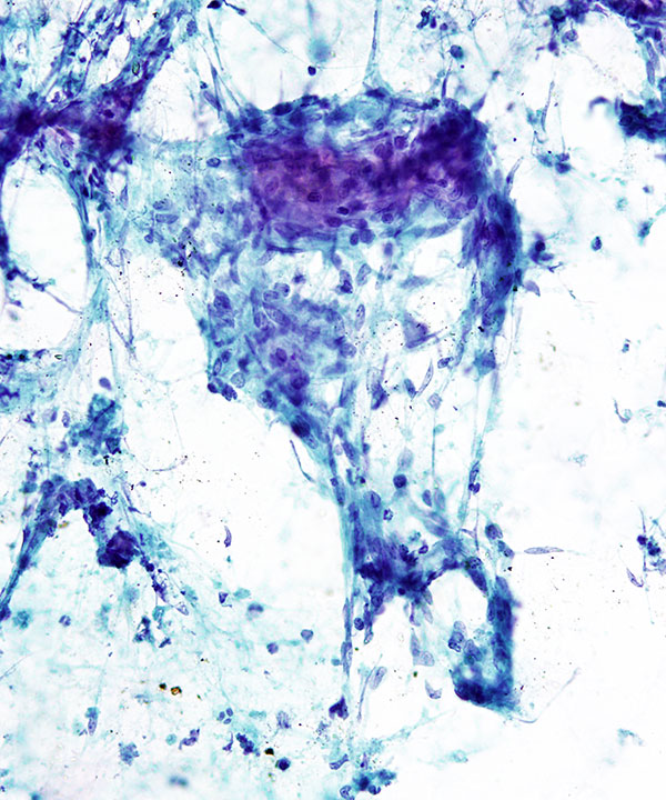
DQ stain: Low power
Lower bottom cohesive clusters with irregular edges
Note scattered giant cells
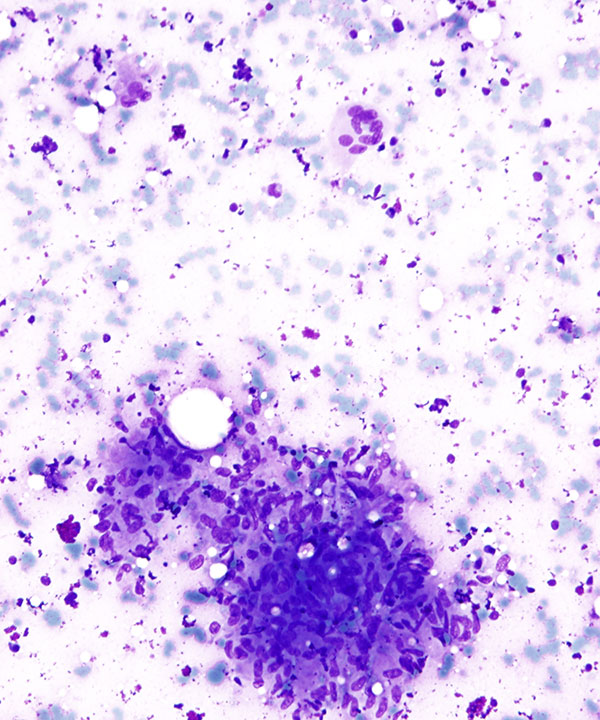
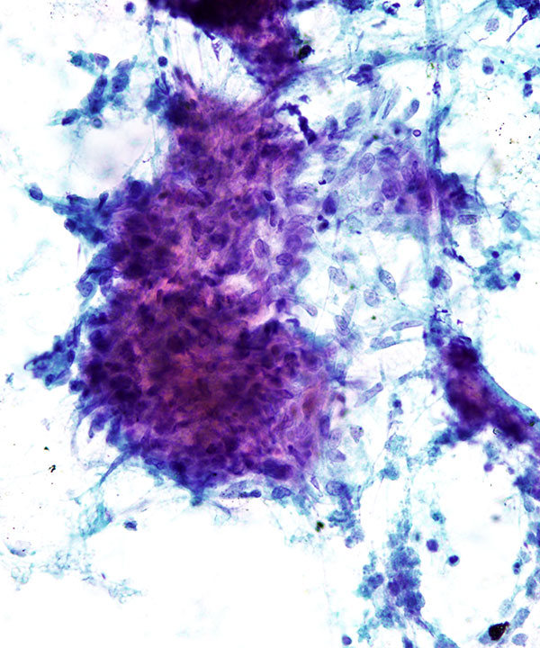
DQ Stain: High power
Cohesive cluster with syncytial appearance, abundant pale blue cytoplasm
Note on right Langhans-type giant cell
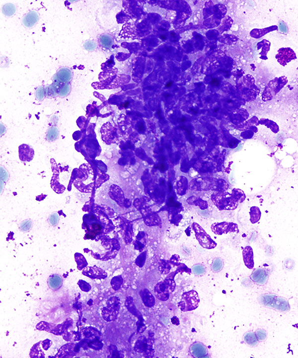
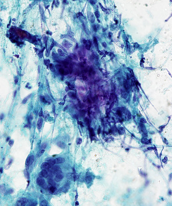
DQ stain: High power
Note spindled/slipper shape of nuclei
Pale blue/frothy cytoplasm and lack of cell membranes
Due to irregular nuclei and overlap of nuclei, may be mistaken for malignancy
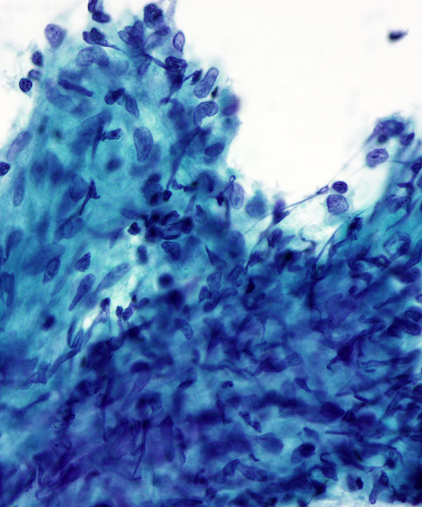
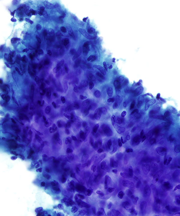
Pap stain: High power
Loose aggregate of epithelioid histiocytes with bean shaped nuclei, dense cytoplasm and lymphocytic crush artifact.
DQ stain: High power
Multinucleated giant cell with eccentrically placed cluster of nuclei (Langhans-type)
Note giant cells are NOT required for the diagnosis of Granulomatous inflammation
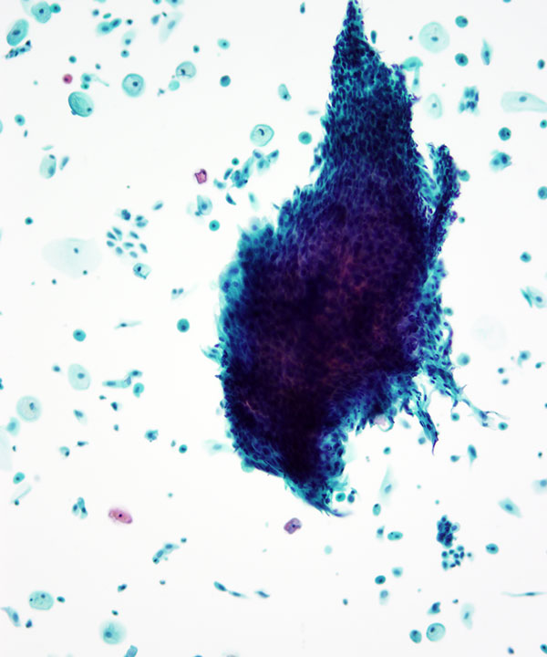
Pap stain: Low power
Liquid based preparation with aggregate of epithelioid histiocytes.
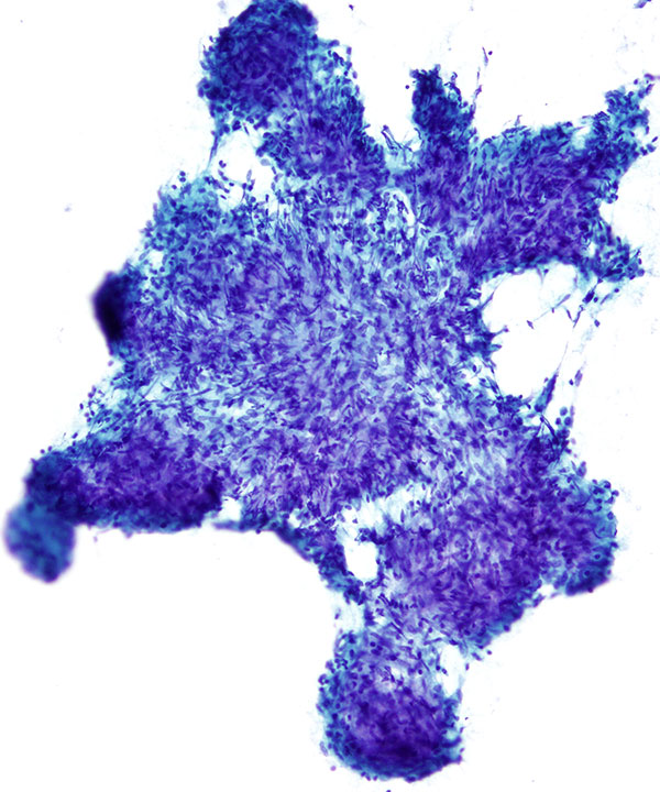
Pap smear: Low power
Note the cytoplasm is pale and indistinct and the cluster has syncytial appearance
Pap stain: High power
Liquid based preparation with aggregate of epithelioid histiocytes. Note dense cytoplasm.
Pap stain; Medium power
Cohesive group of histiocytes with spindled/slipper shaped nuclei (may get mistaken for spindle cell neoplasm) BUT the cells have low N:C ratio and no atypia
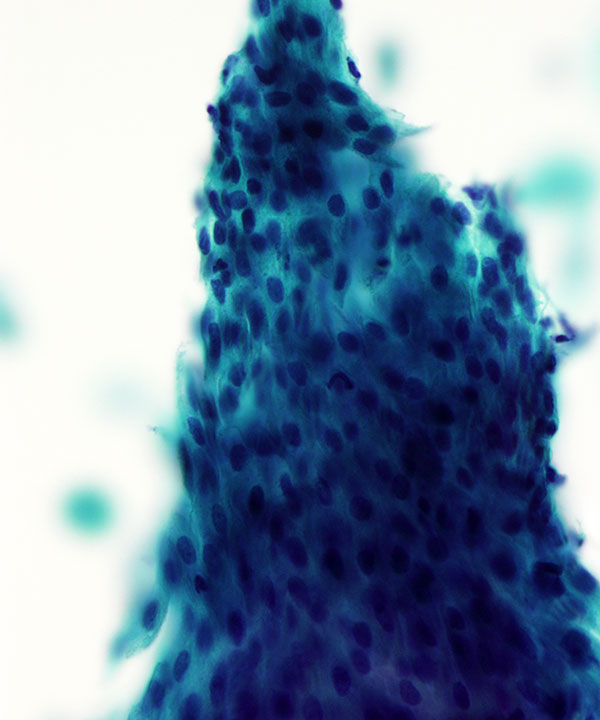
Pap smear: High power
Epithelioid granuloma with background necrosis (upper right and middle)
This would be an example of Necrotizing granuloma
Pap smear: High power
Note the cytoplasm is pale and indistinct and the cluster has syncytial appearance
Langhans-type giant cell on the right
Pap smear: High power
Syncitial cluster of epithelioid histiocytes
Note the cytoplasm is pale and indistinct and the spindled and bean shaped nuclei of the cells
Features
• Variable cellularity
• Predominantly cohesive fragments
• Clustered mononucleated, epithelioid histiocytes (MUST for diagnosis)
• Indistinct cell membranes giving rise to syncytial/squamoid appearance
• Pale blue cytoplasm (DQ) to pale clear cytoplasm (Pap)
• Multinucleated giant cells (NOT required for diagnosis)
• Boomerang/carrot/slipper shaped nuclei
• May seem spindled
• Nuclei may be irregular and show grooves
• N:C ratio are normal
• May be associated with necrosis (Necrotizing granulomas)
• Necrotizing granulomas may be caseating/infectious or non-infectious
• Necrosis may be absent in infections, Sarcoidosis and malignancy associated granulomas
• Recommend requesting microbial cultures (in a comment or during immediate assessment)
• Recommend performing AFB (mycobacteria) and GMS (fungal) stains
• May see negative images of fungal organisms on DQ stain
• With negative stains and no necrosis; DO NOT report as Sarcoidosis (it is a clinical diagnosis)
• With negative stains and cultures may be due to underlying malignancy (recommend excision)
Silverman JF et al. Acta Cytol. 1985 Jul-Aug;29(4):535-41.