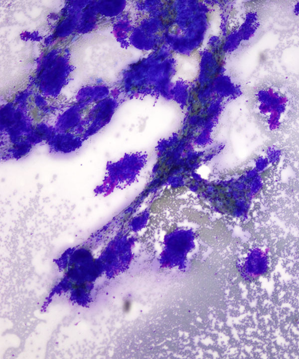
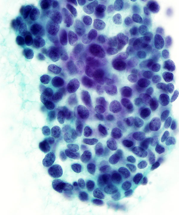
Low power: DQ stain
Cellular smear with small basaloid cells and hyaline material, metachromatic on DQ stain
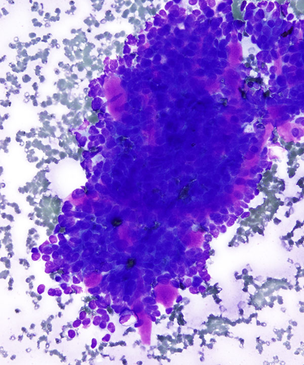
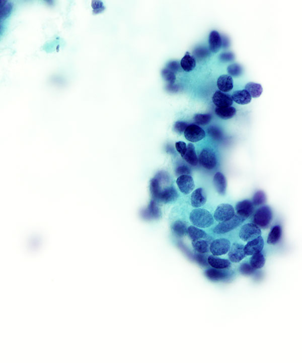
Medium power: DQ stain
Cellular smear with small basaloid cells surrounded by hyalinized stromal material and hyaline globules, metachromatic on DQ stain
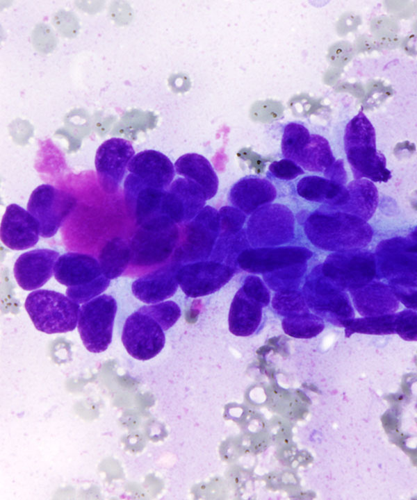
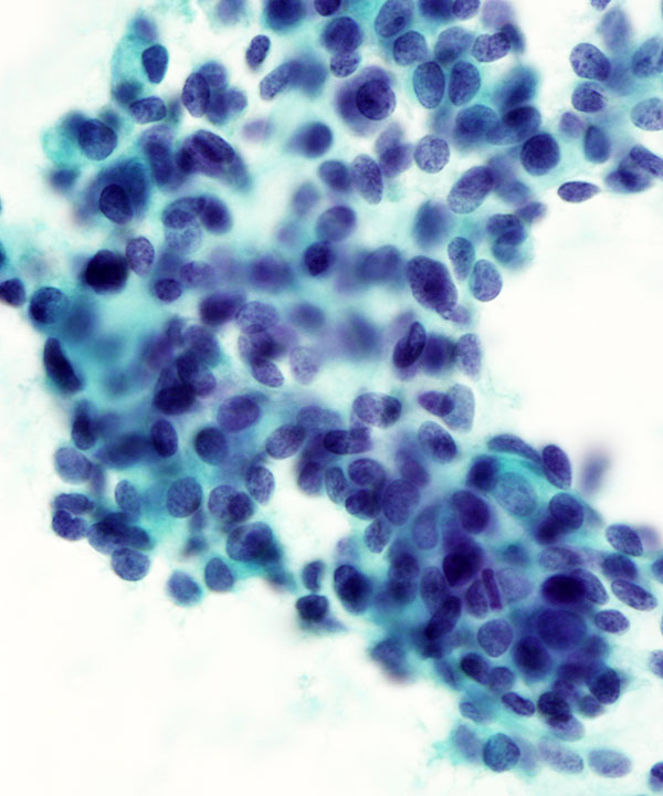
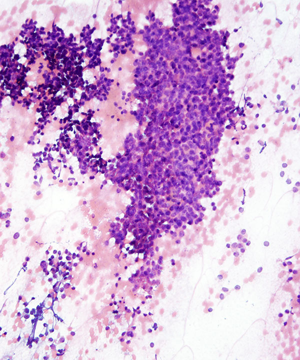
Medium power: Pap stain
Cellular smear with small basaloid cells surrounding hyalinized stromal material, pale translucent on Pap stain
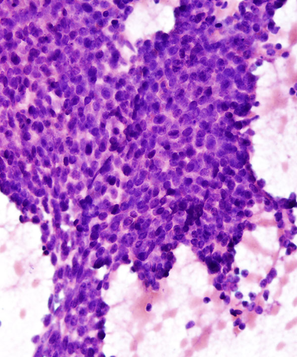
High power: Pap stain
Small basaloid cells with round uniform nuclei containing slightly coarse chromatin and small nucleoli. Pale, translucent hyalinized stroma can also be appreciated.
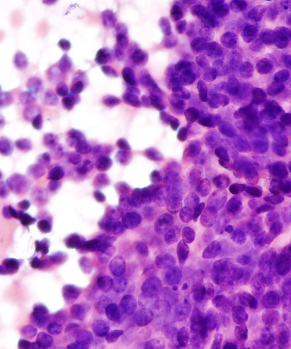
High power: Pap stain
Small basaloid cells with round uniform nuclei containing slightly coarse chromatin and small nucleoli. Pale, translucent hyalinized stroma can also be appreciated.
High power: Pap stain
Small basaloid cells with round uniform nuclei containing slightly coarse chromatin and small nucleoli. Pale, translucent hyalinized stroma can also be appreciated.
High power: Pap stain
Small basaloid cells with round uniform nuclei containing slightly coarse chromatin and small nucleoli. Pale, translucent hyalinized stroma can also be appreciated.
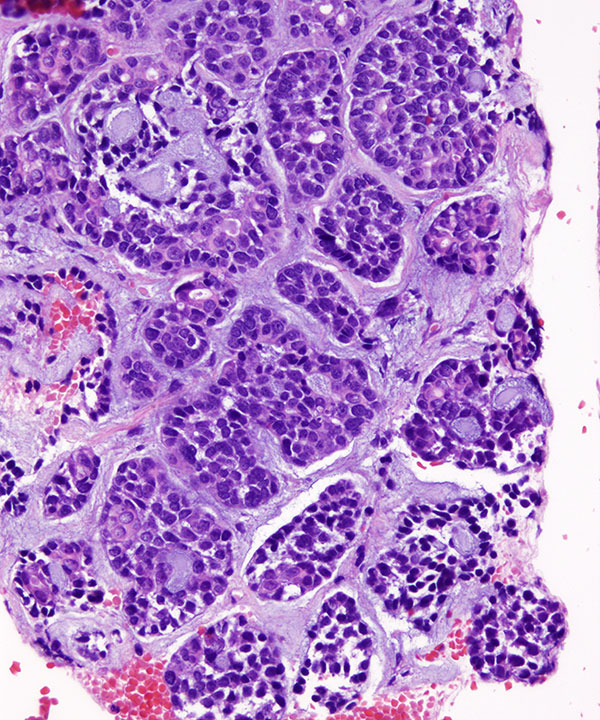
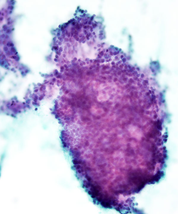
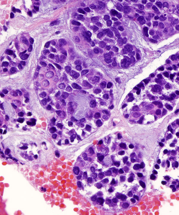
Cell block: H&E stain
Cribriform arrangement of adenoid cystic carcinoma
Cell block: H&E stain
Small uniform basaloid cells with cribriform arrangement
Medium power: H&E stain
Solid variant of adenoid cystic carcinoma
Also called poorly differentiated adenoid cystic carcinoma
Cellular smears with solid mass of basaloid cells
Note lack of hyaline material
High power: H&E stain
Solid variant of adenoid cystic carcinoma
Solid mass of basaloid cells with nuclear enlargement, coarse chromatin and lack of hyaline material
High power: H&E stain
Solid variant of adenoid cystic carcinoma
Basaloid cells with nuclear enlargement, coarse chromatin and lack of hyaline material
D/D includes other basaloid/small cell tumors
Features
• Cellular smears
• Small basaloid cells
• Cribriform, rosettes, tubular clusters
• Small, uniform, relatively bland basaloid cells
• Scant cytoplasm
• Cells surrounded by hyalinized stroma
• Cells are arranged around pseudoglandular spaces containing basement membrane like material
• Nuclei small, round to oval, rare atypia
• Chromatin may be coarse
• Rare visible small nucleoli
• Numerous naked nuclei
• Metachromatic homogenous hyaline material: globules or cylinders
• Rare mitosis or necrosis
• Important to differentiate from pleomorphic adenoma where the hyaline material is more fibrillar, spindle cells infiltrating the stroma and lack of naked nuclei in the background. Also difficult to differentiate other basaloid tumors like basal cell adenoma/carcinoma and epithelial myoepithelial carcinoma; therefore unequivocal diagnosis of adenoid cystic carcinoma is only given where there is definitive clinical evidence of malignancy
• Express cytokeratin, vimentin, actin, CEA, CD117
• t(6;9) can be seen
Jayaram N et al. Diagn Cytopathol. 1989;5(4):349-54.
Qizilbash AH et al.Acta Cytol. 1985 Jul-Aug;29(4):503-12.
Klijanienko J et al.Diagn Cytopathol. 1997 Jul;17(1):36-41.
Fehr A, et al. J Pathol. 2011 Jul;224(3):322-7.