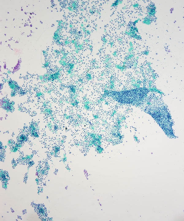
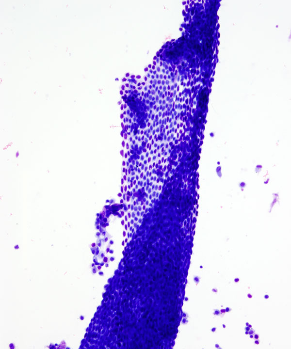
Low power: Pap stain
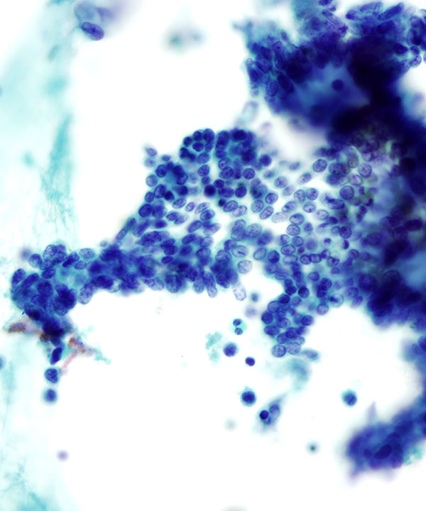
Pelvic washing with flat sheets of benign mesothelial cells in a background of scattered mesothelial cells, histiocytes and inflammatory cells.
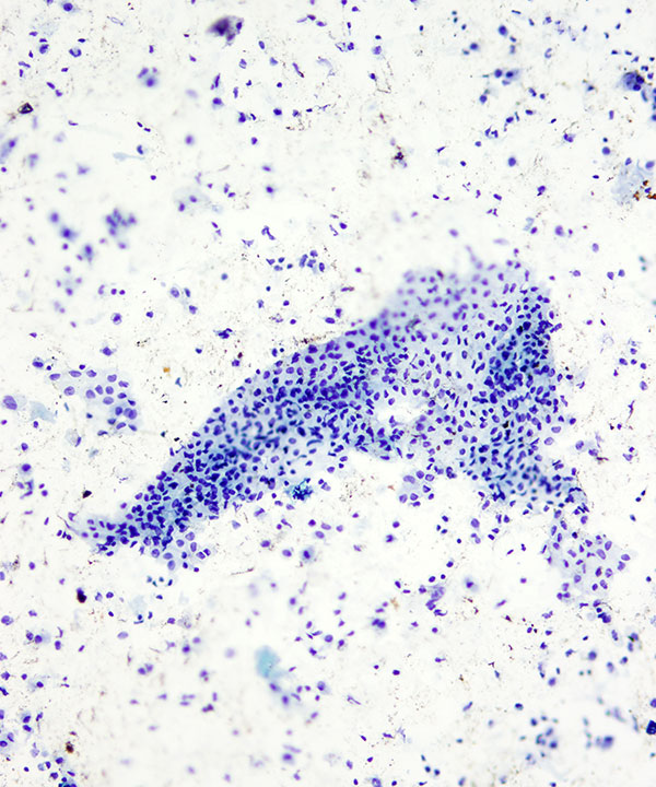
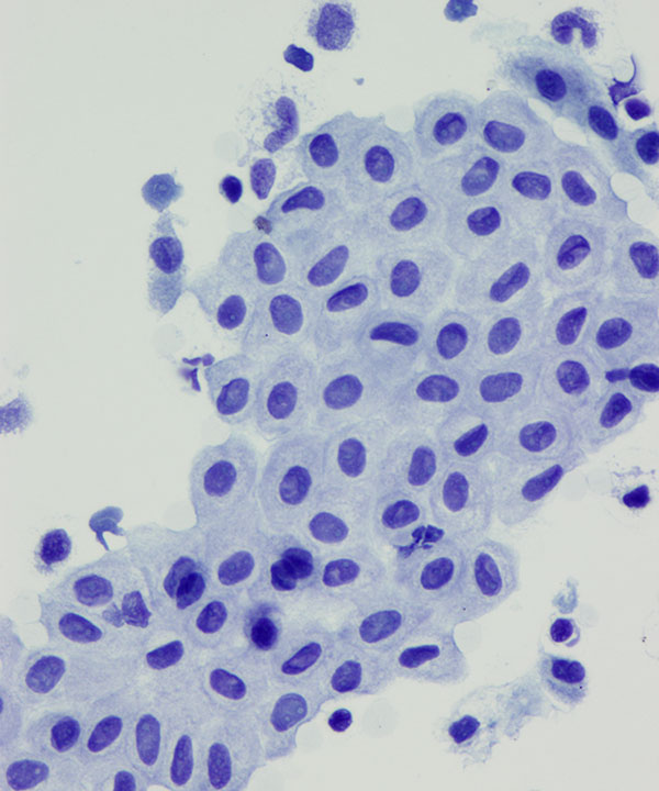
Low power: Pap stain
Flat sheet of mesothelial cells with round to oval nuclei, smooth to slightly irregular nuclear membranes, and dense cytoplasm with windows between cells
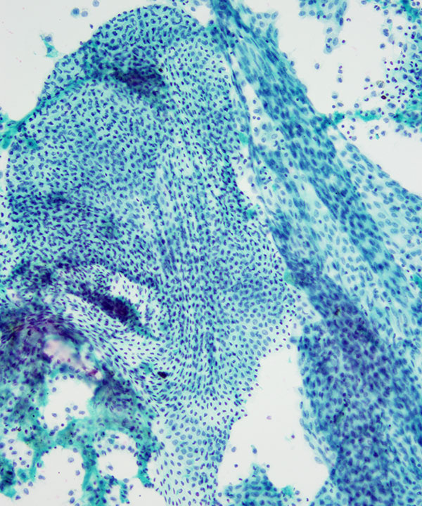
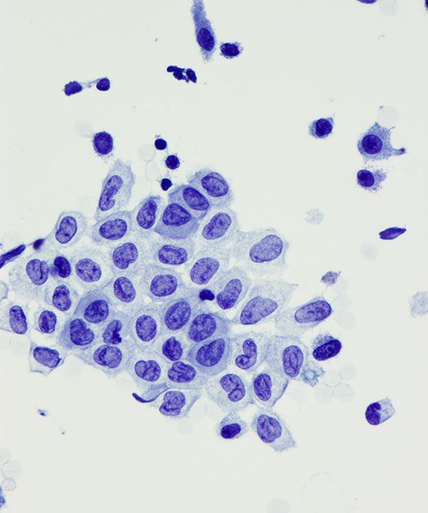
Medium power: Pap stain
Flat sheet of benign mesothelial cells, some with reactive changes
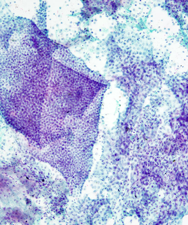
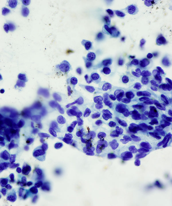
Medium power: Pap stain
Pelvic washing with flat sheets of benign mesothelial cells in a background of scattered mesothelial cells, histiocytes and inflammatory cells
Note how the flat sheets fold and roll over on themselves
Medium power: Pap stain
Flat sheets of benign mesothelial cells folded over with evenly spaced out cells
Medium power: Pap stain
Mesothelial cells in a washing with flat sheets of cells
Medium power: Pap stain
Flat sheet of mesothelial cells with round to oval nuclei, smooth to slightly irregular nuclear membranes, and dense cytoplasm with windows between cells
Note inflammatory cells in the background
Medium power: Pap stain
Flat sheet of mesothelial cells in a washing.
Note the wrinkled nuclei with irregular nuclear membranes often seen in washings
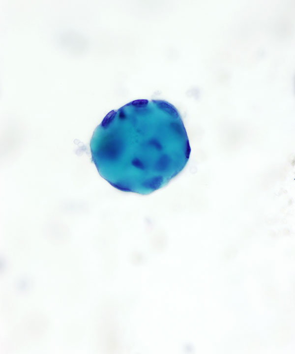
High power: Pap stain
Spherical collagen ball surrounded by benign flattened mesothelial cells, commonly seen in washings
Medium power: Pap stain
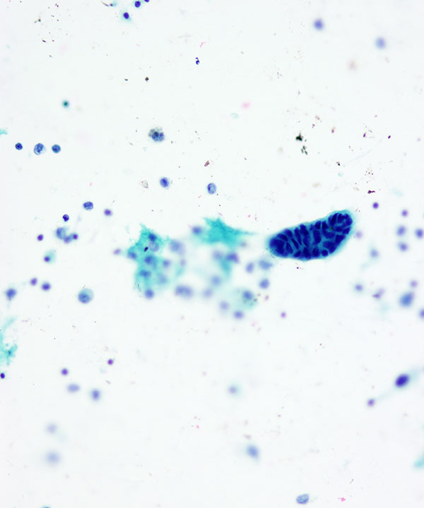
Benign glandular elements of Mullerian origin. Sometimes cilia may be present indicative of tubal elements.
Most washings are obtained as part of a surgical procedure with a corresponding histologic specimen. It might be advisable to correlate findings with the histologic specimen as these often result in false positive interpretations of washings.
Medium power: Pap stain
Benign glandular elements of Mullerian origin
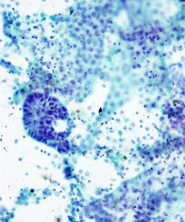
Medium power: Pap stain
Glandular elements of Mullerian origin. Often requiring correlation with histologic specimen to rule out involvement by serous borderline tumors with minimal atypia.
Features
• Typically obtained as part of cancer staging procedure (primarily gynecologic but also others like gastric or pancreaticobiliary). Also done to rule out malignant involvement and assessing response to treatment.
• Mesothelial cells predominantly in flat sheets
• Rarely single cells
• Evenly spaced uniform cells
• Cells often have windows (space between cells)
• Moderate to abundant cytoplasm
• Round to oval nuclei
• Nuclear membrane often irregular and wrinkled
• Chromatin usually fine
• Small nucleoli, may be prominent when reactive
• Histiocytes, single or in aggregates
• Inflammatory cells
• May have:
- Psammoma bodies (benign conditions like endosalpingiosis, ovarian cystadenomas, as well as malignancies e.g. ovarian carcinoma, papillary carcinomas and mesotheliomas)
- Calcification
- Collagen balls (benign and malignant conditions)
- Benign glands of endosalpingiosis (may have ciliated fallopian tube-like epithelium)
- Benign glands of endometrial glands (may have hemosiderin laden macrophages suggestive of endometriosis)