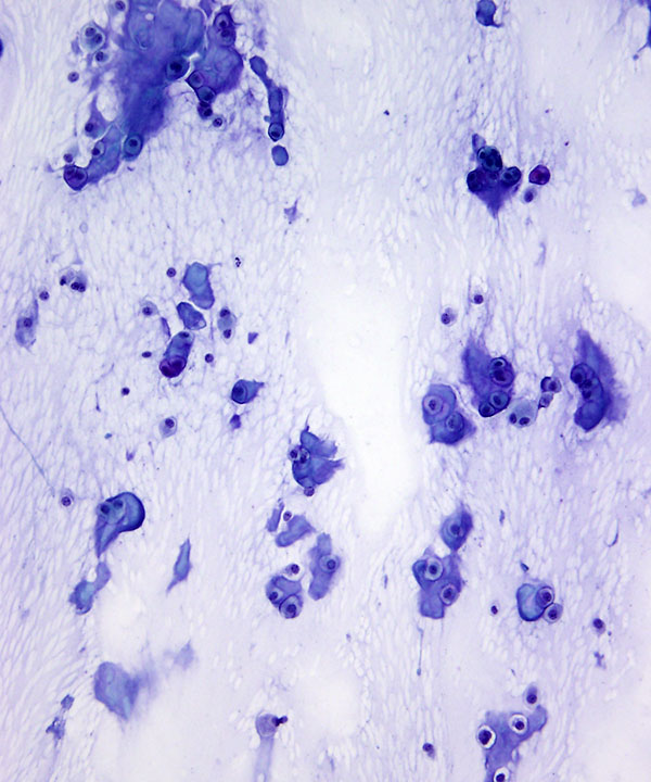
Low Power; Pap Stain
Moderately cellular smear, fragments of hyaline cartilage and thin chondromyxoid background
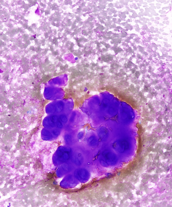
High Power; DQ stain
Magenta colored fragment of hyaline cartilage. The fragment shows increased cellularity
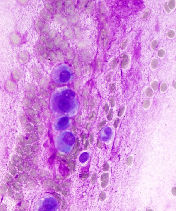
High Power; DQ stain
Single neoplastic cells dispersed in thin myxoid background
Rare cells with binucleation
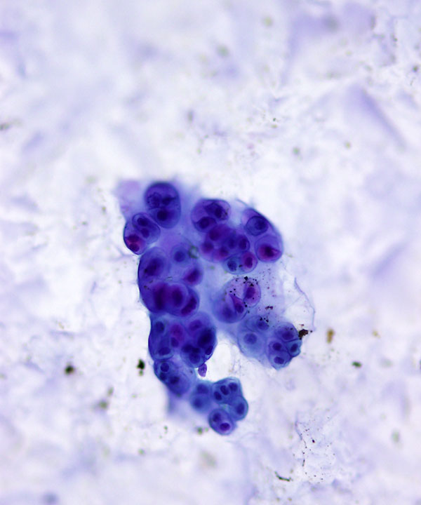
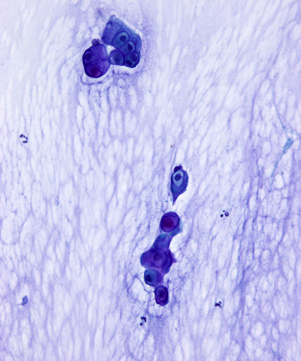
High Power; Pap stain
Hyaline cartilage fragment with high cellularity
Cells have distinct cell borders, hyperchromatic nuclei and clear cytoplasm
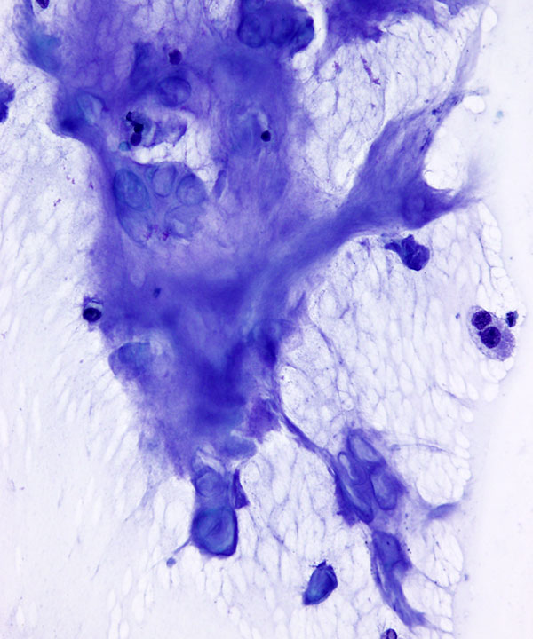
High Power; Pap stain
Fragment of metachromatic matrix with myxoid background and hyaline cartilage
High Power; Pap stain
Hyaline cartilage fragment and single cells with hyperchromatic nuclei, clear cytoplasm and prominent cell borders.
Background shows thin myxoid matrix
Features
• Low to moderate cellularity
• Cohesive fragments of hyaline cartilage (Appear magenta on DQ stain, and clear on Pap stain)
• Variable cellularity of cartilage fragments
• Rare single cells
• Cells located in lacuna
• May have more than one cell per lacuna
• Distinct cell borders and clear cytoplasm
• Eccentrically placed nuclei
• Round nuclear contours
• Condense chromatin
• Single small nucleolus
• Occasional binucleated cells
• Minimal to no atypia, pleomorphism
• May have thin myxoid/chondromyxoid background
• No mitoses, no necrosis
• Diagnosis requires compatible radiologic findings
• Morphology is indistinguishable from low grade chondrosarcoma
• Recommend reporting as Low Grade Chondroid Neoplasm; comment on radiologic correlation
Thool A et al . Acta Cytol. 2001 Jan-Feb;45(1):86-8.