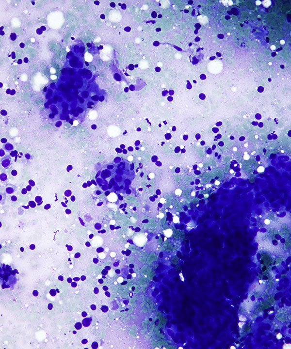
Low power: DQ stain
Cellular smear with 3D crowded clusters and numerous singly dispersed malignant cells with enlarged round to oval to irregular nuclei, occasional prominent nucleoli.
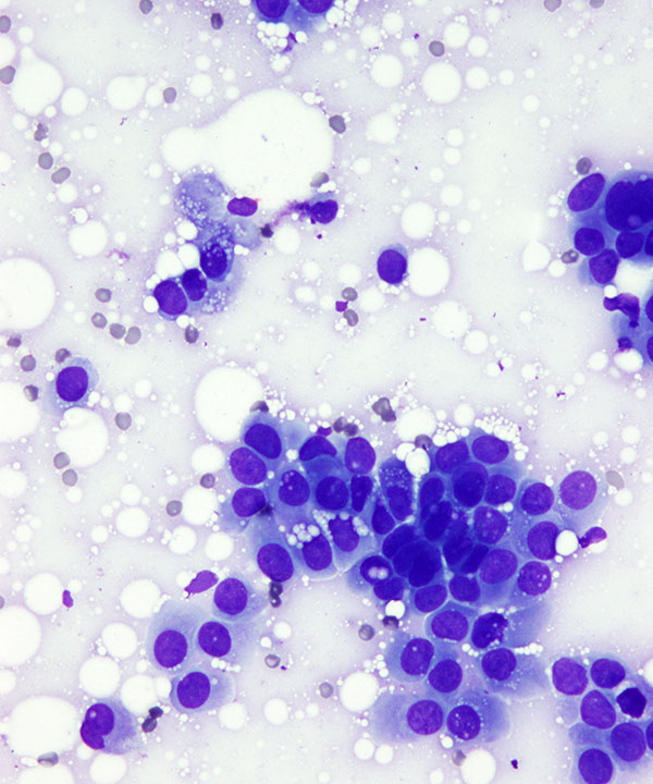
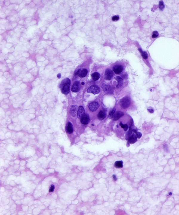
High power: DQ stain
3D cluster of malignant cells, enlarged round to oval to irregular nuclei, occasional prominent nucleoli. Note plasmacytoid appearance with eccentric nuclei and the presence of single cells in the background. Note intracytoplasmic lipid that dissolves on processing leaving multiple small cytoplasmic vacuolations.

Low power: Pap stain
Loosely cohesive 3D clusters and single malignant cells with enlarged round to oval to irregular nuclei, occasional prominent nucleoli. Note cellular molding and apoptotic cells. Single cells have a plasmacytoid appearance.
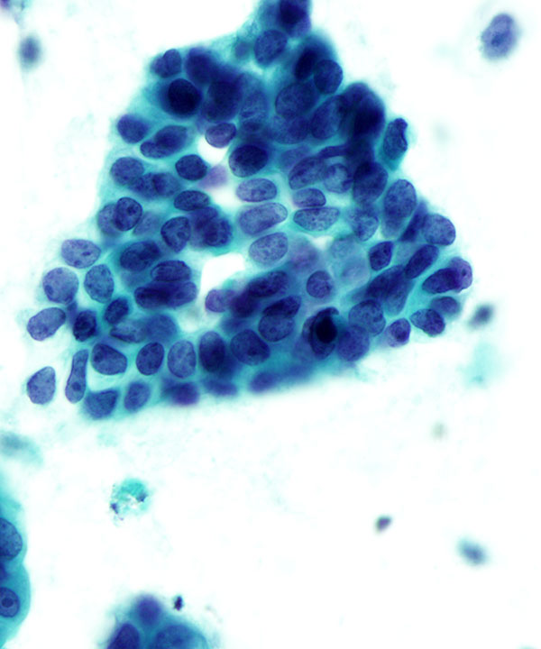
High power: Pap stain
3D cluster of malignant cells, enlarged round to oval to irregular nuclei, occasional prominent nucleoli. Note cellular molding and apoptotic cells. Note presence of single cells in the background.
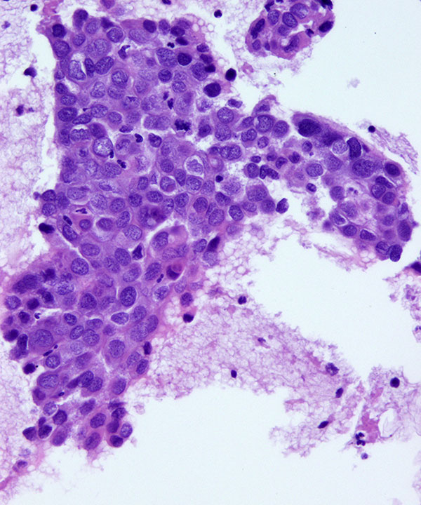
Cell block H&E stain
Sheet of malignant cells with enlarged irregular nuclei, intracytoplasmic lumen with mucin and mitotic figures.
Cell block H&E stain
Small acinar cluster of malignant cells. Note intracytoplasmic lumen with mucin.
Features
• Cellular smears
• Disorganized sheets and single cells
• 3D crowded clusters
• Microacini
• May have single cell filing
• Absence of myoepithelial cells
• Plasmacytoid appearance
• Moderate amounts of vacuolated cytoplasm
• Intracytoplasmic lumens with mucin (targetoid mucin vacuoles)
• Intracytoplasmic lipid (shows as mutiple small vacuolations)
• Enlarged nuclei with nuclear irregularity
• Coarse chromatin
• Conspicuous to prominent nucleoli
• May have necrosis
• May have squamous, apocrine or clear cell metaplasia
• Calcifications, Mitotic figures, Apoptosis
• Express cytokeratin, CK7, CK8, CK18, GATA3, may express mammaglobin, GCDFP-15, ER, PR, E-Cadherin
Wang HH et al. Am J Clin Pathol. 1989 Dec;92(6):736-40.
Mouriquand J et al.Acta Cytol. 1980 Mar-Apr;24(2):153-9.