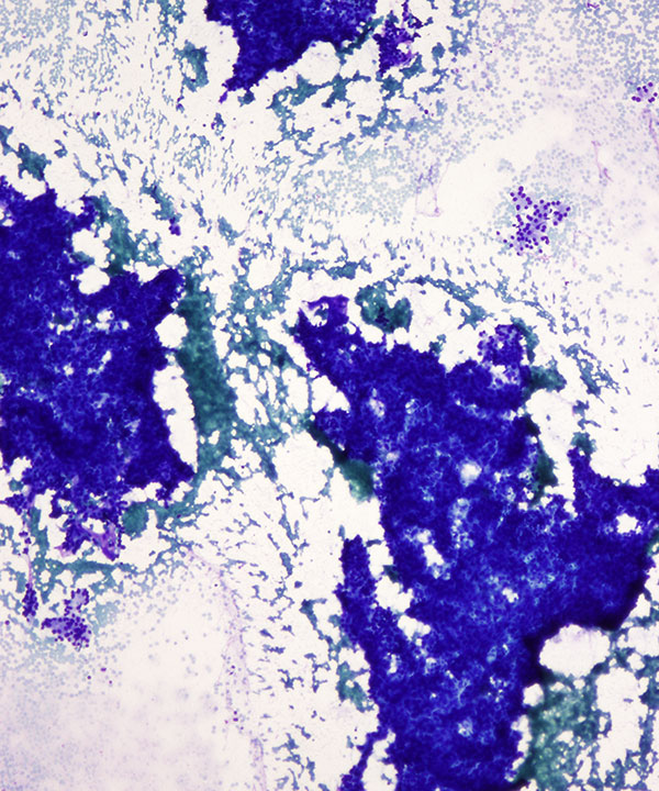
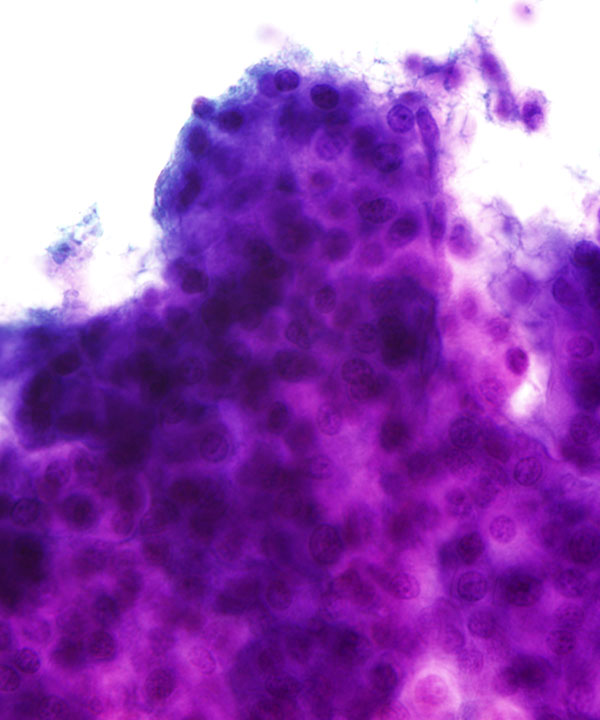
Low power: DQ stain
Cellular smear of hepatocytes in 3D aggregates. Note a few small groups showing acinar-like features
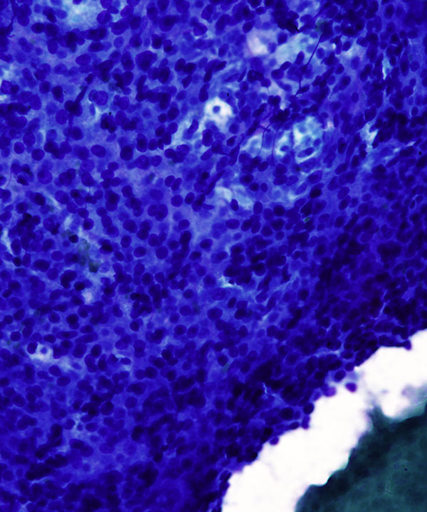
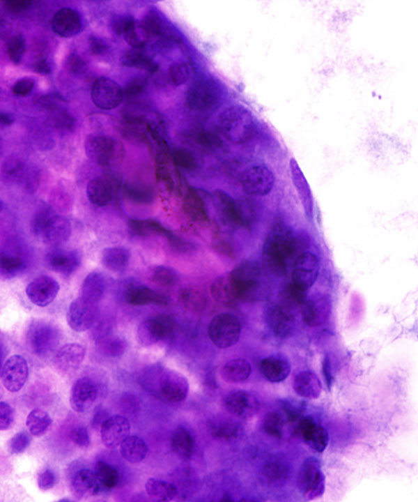
Medium power: DQ stain
3D smear of hepatocytes with thickened trabeculae and transgressing capillaries
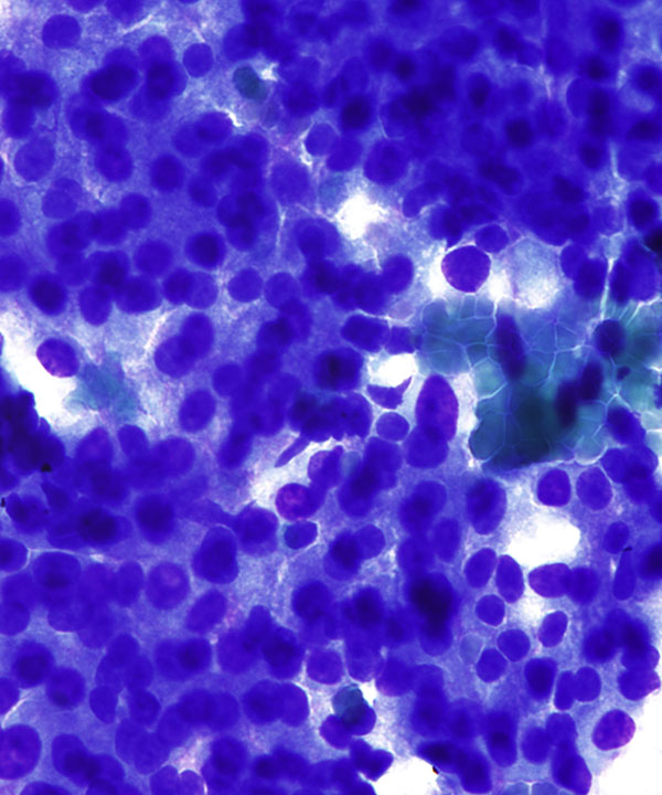
High power: DQ stain
Hepatocytes with higher N:C ratios, round to oval nuclei and granular cytoplasm
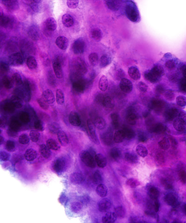
Medium power: Pap stain
Hepatocytes with trangressing capillaries
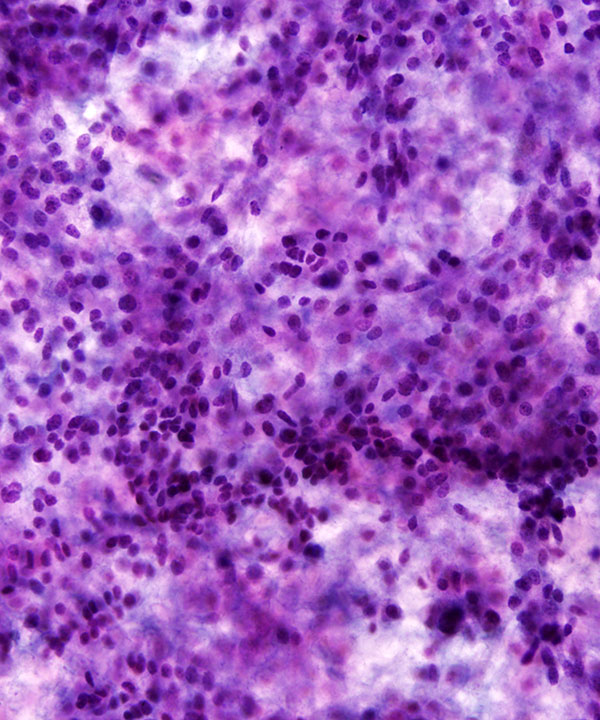
High power: Pap stain
Hepatocytes cell groups with endothelial cell wrapping
High power: Pap stain
Hepatocyte cell groups with endothelial cell wrapping
Note hepatocytes with round slightly irregular nuclei, coarse chromatin and granular cytoplasm
High power: Pap stain
Hepatocytes with round to slightly irregular nuclei, coarse chromatin, visible nucleoli and granular cytoplasm
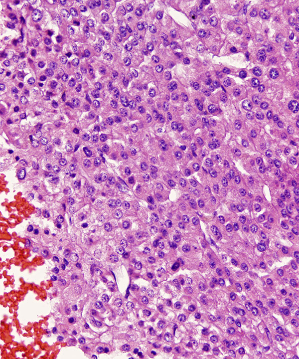
Cell block: H&E stain
Malignant cell groups with thickened cell plates and endothelial wrapping
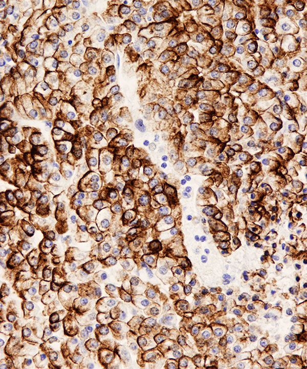
Cell block: Cam5.2 stain
Diffuse membranous staining in malignant cells
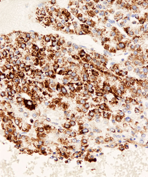
Cell block: HepPar1 stain
Granular cytoplasmic staining in malignant cells
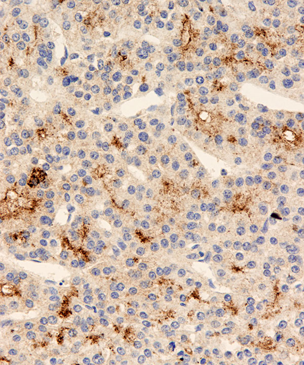
Cell block: Glypican 3 stain
Granular cytoplasmic staining in malignant cells
Features
• Highly cellular smears
• Thickened trabeculae (3 to >10 cell thick)
• Groups of cells with endothelial cell wrapping
• May have crowded 3D groups
• May have acinar structures
• Capillaries traversing cell groups
• Cells resemble normal hepatocytes
• Round to polygonal cells
• May have increased N:C ratios
• Granular cytoplasm
• Well defined borders
• May have bile in cytoplasm or in canaliculi
• May have cytoplasmic hyaline globules (alpha-anti trypsin or alpha-fetoprotein), or Mallory bodies
• Nuclei usually round, may be enlarged
• Nuclei central to eccentrically located
• Variation in nuclear size may be seen
• Frequent bi/multinucleation
• Naked nuclei present
• May have satellite nuclei around nucleus
• Prominent nucleoli, may be multiple
• Usually clean background
• Necrosis typically not present
• May have mitosis
• Usually bile duct cells not present
• Lipofuscin and iron usually not present
• Positive for CK8/CK18, HepPar-1, Glypican-3 (D/D benign or adenoma), Arginase-1, canalicular pattern of p-CEA and CD10, cytoplasmic TTF-1; usually negative for cytokeratin, EMA, MOC-31, CK7, CK20, CK19
Tao LC et al.Acta Cytol. 1979 Jul-Aug;23(4):287-91.