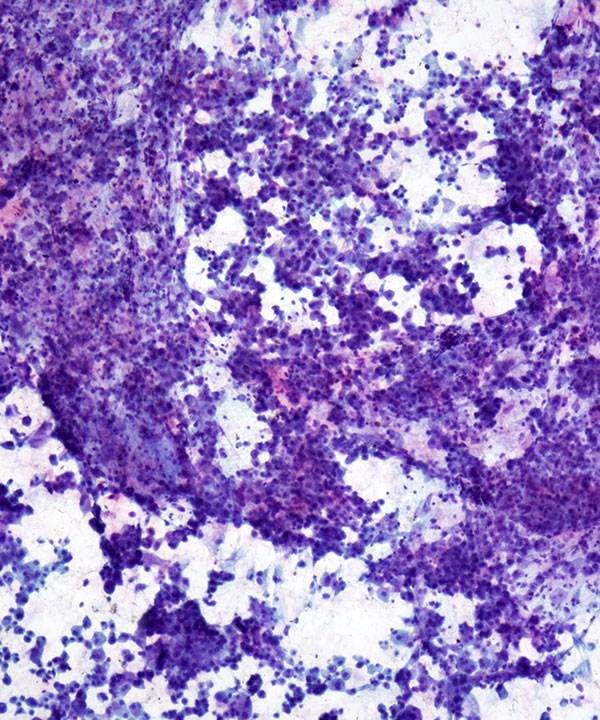
Low power: Pap stain
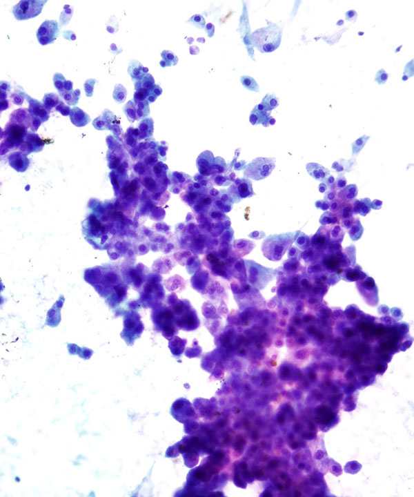
Cellular smear with small groups and single malignant cells
Low power: Pap stain
Nests of malignant large cells with nuclear atypia, frequent binucleation, inconspicuous nucleoli, and granular cytoplasm
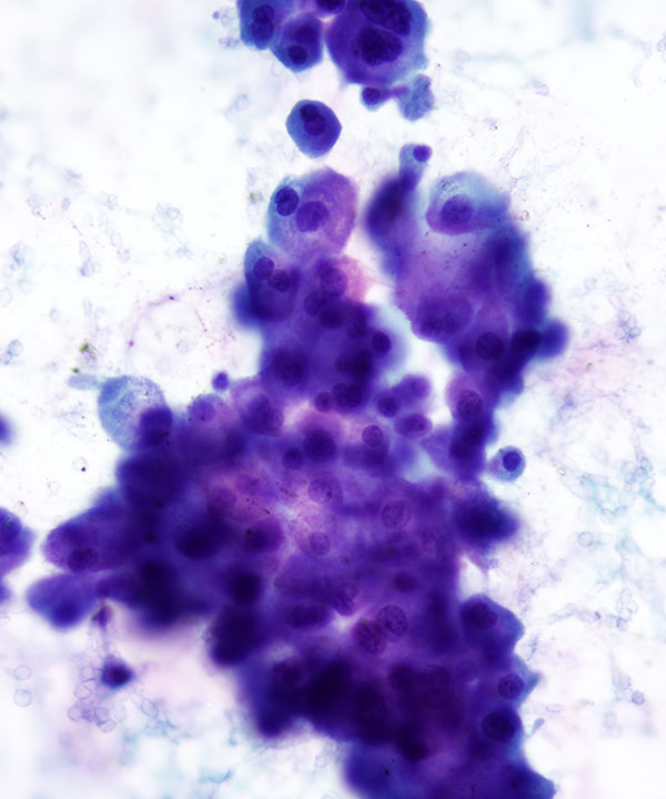
High power: Pap stain
Malignant large cells with enlarged, wrinkled nuclei, frequent binucleation, inconspicuous nucleoli, and granular cytoplasm
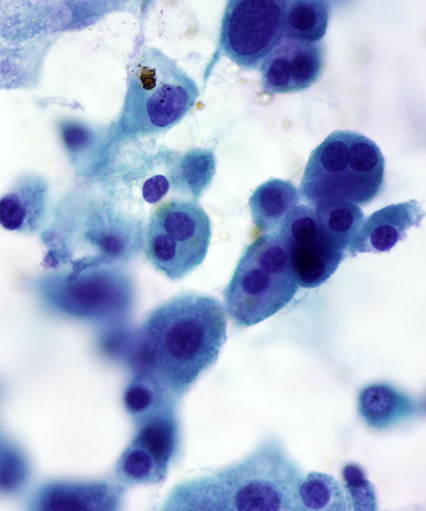
High power: Pap stain
Malignant large cells with nuclear atypia, binucleation, inconspicuous nucleoli, and granular cytoplasm.
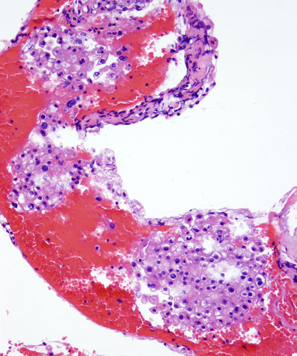
Cell block: H&E stain
Solid nests of chromophobe RCC cells with nuclear atypia and granular cytoplasm.
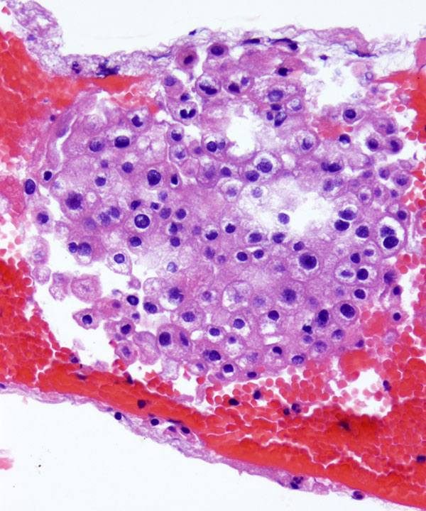
Cell block: H&E stain
Solid nests of malignant cells with koilocytic nuclei, binucleation and granular cytoplasm.
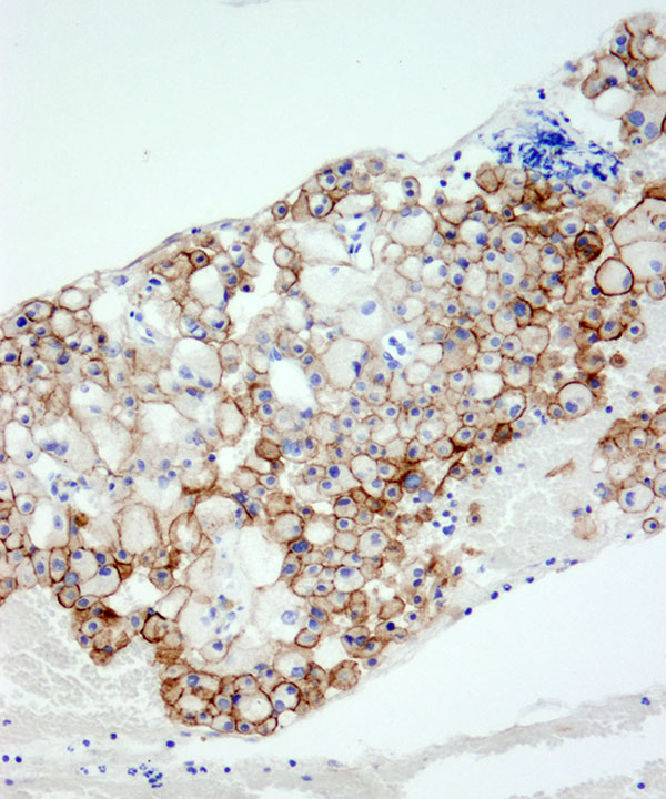
CD117 (c-kit) positive in Chromophobe RCC
Features
• Cellular smears
• Small groups or single cells
• Koilocyte• like cells
• Large polygonal cells
• Abundant pale to granular cytoplasm
• Distinct cell borders
• Enlarged wrinked (raisinoid) nuclei
• Perinuclear clearing (koilocytic)
• Binucleation frequently present
• May have intranuclear cytoplasmic inclusions
• Small inconspicuous nucleoli
• Cytoplasmic microvesicles with acid mucopolysaccharides stain strongly and diffusely blue with Hale colloidal iron (HCI) stain. Differentiate from oncocytoma where HCI is mostly focal.
• Express CD117, EMA, cytokeratin, CK7, RCC+/- , CD10 focal or negative, negative for AMACR and vimentin (D/D clear cell ca)
• Loss of chromosome 1,2,6,10, 13, 17, 21; hypodipolid
Akhtar M et al. Diagn Cytopathol. 1995 Nov;13(4):287-94.